Vertebrae Labeling Worksheet
The Vertebrae Labeling Worksheet is designed for anatomy enthusiasts and students studying the skeletal system. This educational resource provides a clear and comprehensive layout to help users practice identifying and labeling the different vertebrae in the human spine.
Table of Images 👆
- Unlabeled Vertebral Column Worksheet
- Anatomy of the Spine Labeling Worksheet
- Medical School Spine and Vertebrae Labeling Sheet
- Chiropractic Training Vertebrae Diagram Worksheet
- Skeletal System Vertebrae Learning Worksheet
- Detailed Spine Labeling Worksheet for Biology
- Vertebrae Anatomy Study Guide for Students
- Human Spine Structure Labeling Worksheet
- Spinal Cord and Vertebrae Educational Worksheet
- Interactive Vertebrae Structure Labeling Quiz
- Spinal Bones Anatomy Labeling Worksheet
- Printable Vertebrae Identification Activity Sheet
- Human Spine Anatomy Labeling Worksheet
- Spinal Cord and Vertebrae Labeling Worksheet
- Anatomy of Vertebrae Labeling Worksheet
- Vertebrae Identification Labeling Worksheet
- Anatomy Class Vertebrae Labeling Practice
 17 Images of Vertebrae Labeling Worksheet
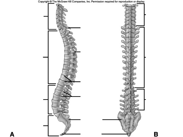
 Unlabeled Vertebral Column Worksheet
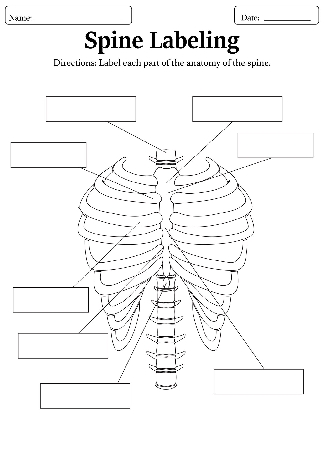
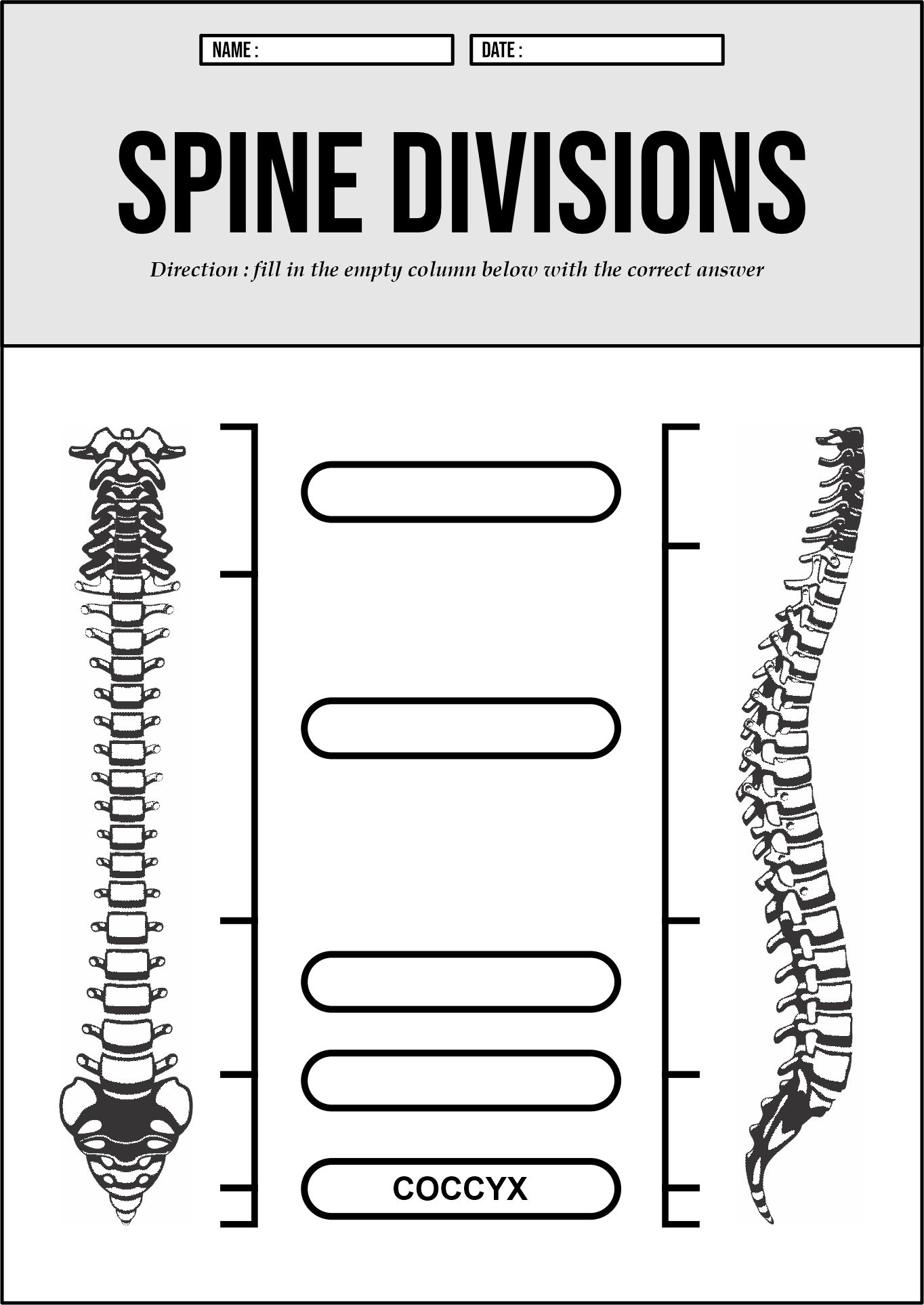
 Anatomy of the Spine Labeling Worksheet
 Medical School Spine and Vertebrae Labeling Sheet
 Chiropractic Training Vertebrae Diagram Worksheet
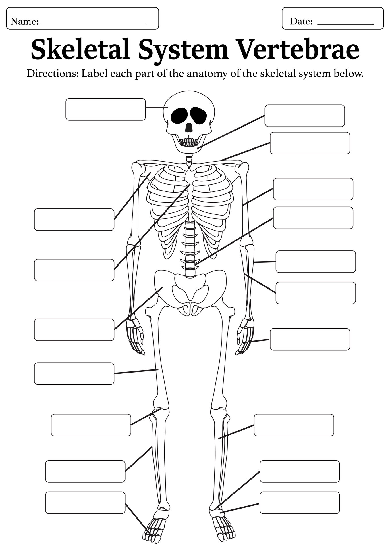
 Skeletal System Vertebrae Learning Worksheet
 Detailed Spine Labeling Worksheet for Biology
 Vertebrae Anatomy Study Guide for Students
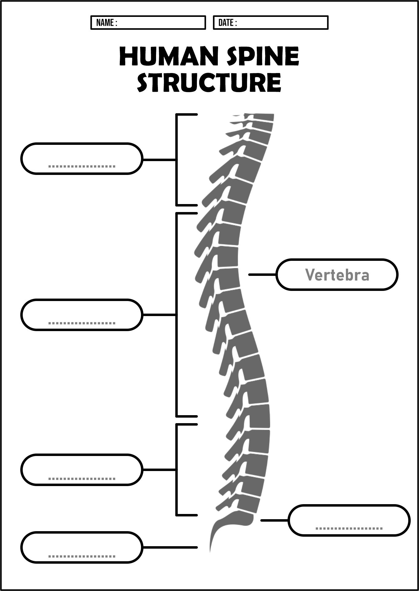
 Human Spine Structure Labeling Worksheet
 Spinal Cord and Vertebrae Educational Worksheet
 Interactive Vertebrae Structure Labeling Quiz
 Spinal Bones Anatomy Labeling Worksheet
 Printable Vertebrae Identification Activity Sheet
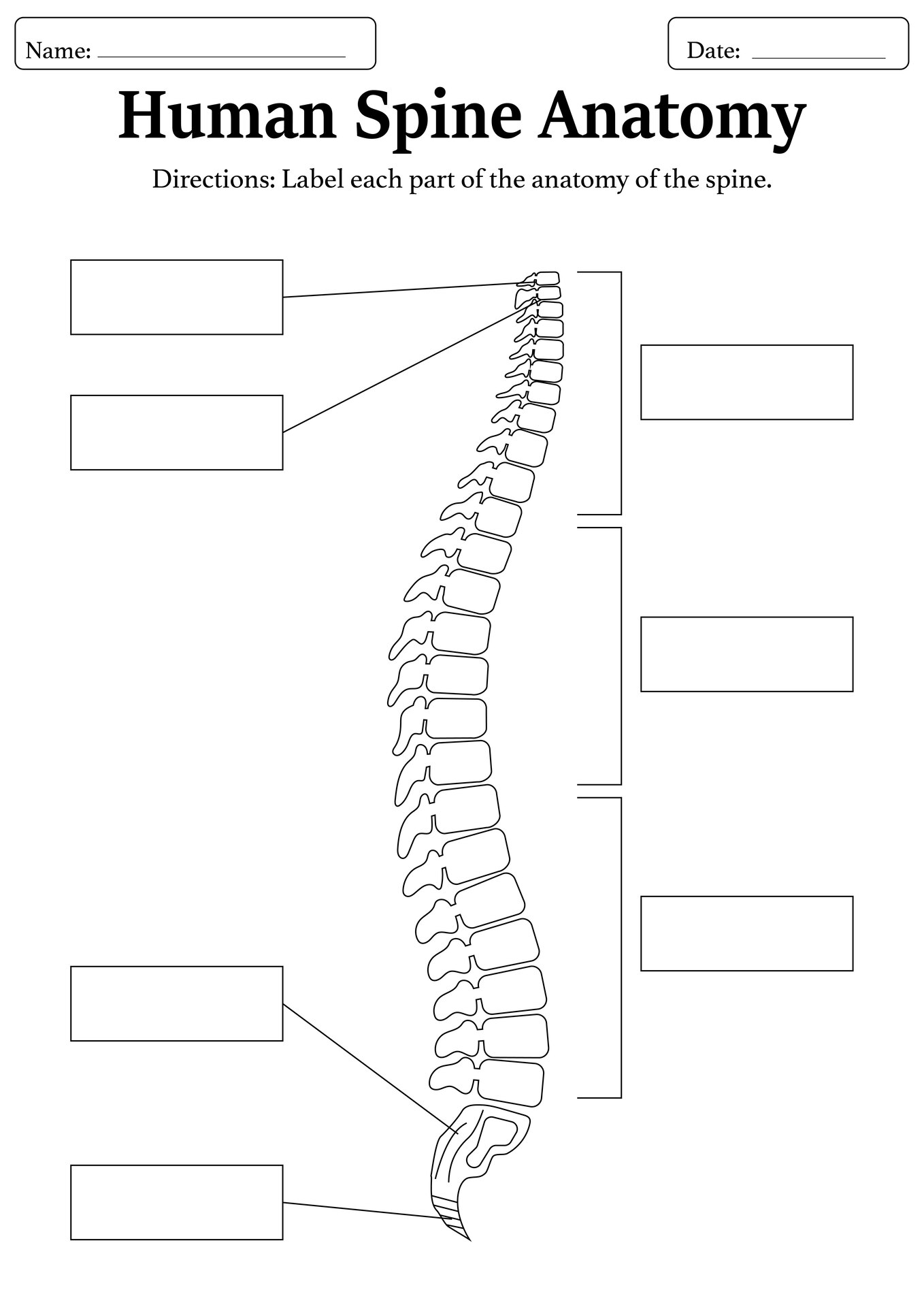
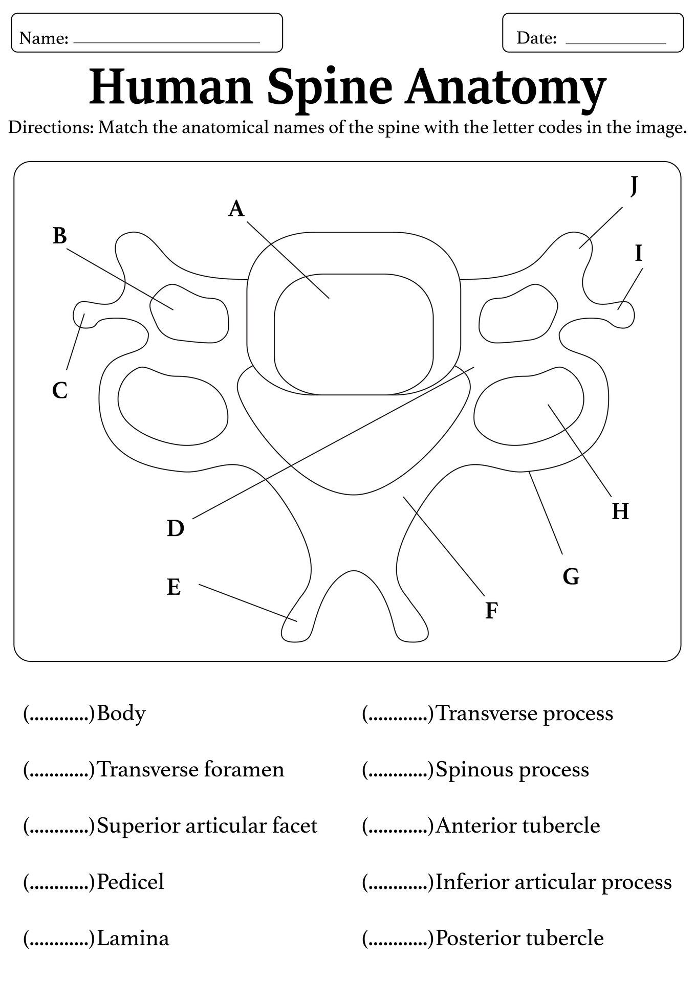
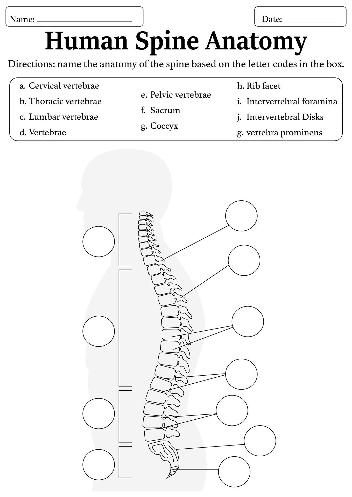
 Human Spine Anatomy Labeling Worksheet
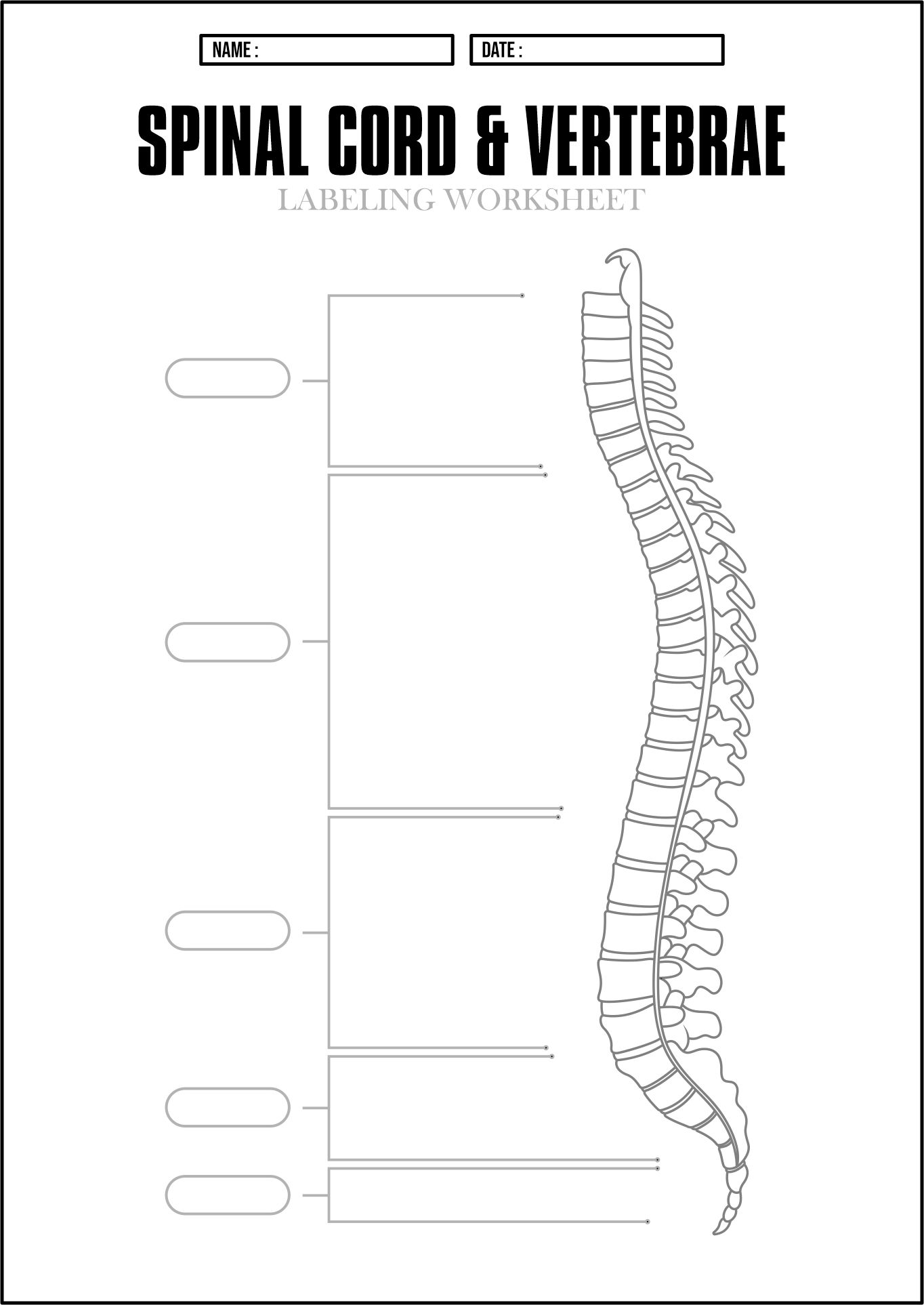
 Spinal Cord and Vertebrae Labeling Worksheet
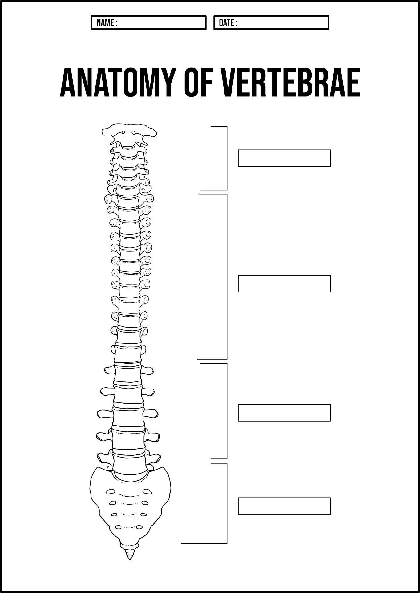
 Anatomy of Vertebrae Labeling Worksheet
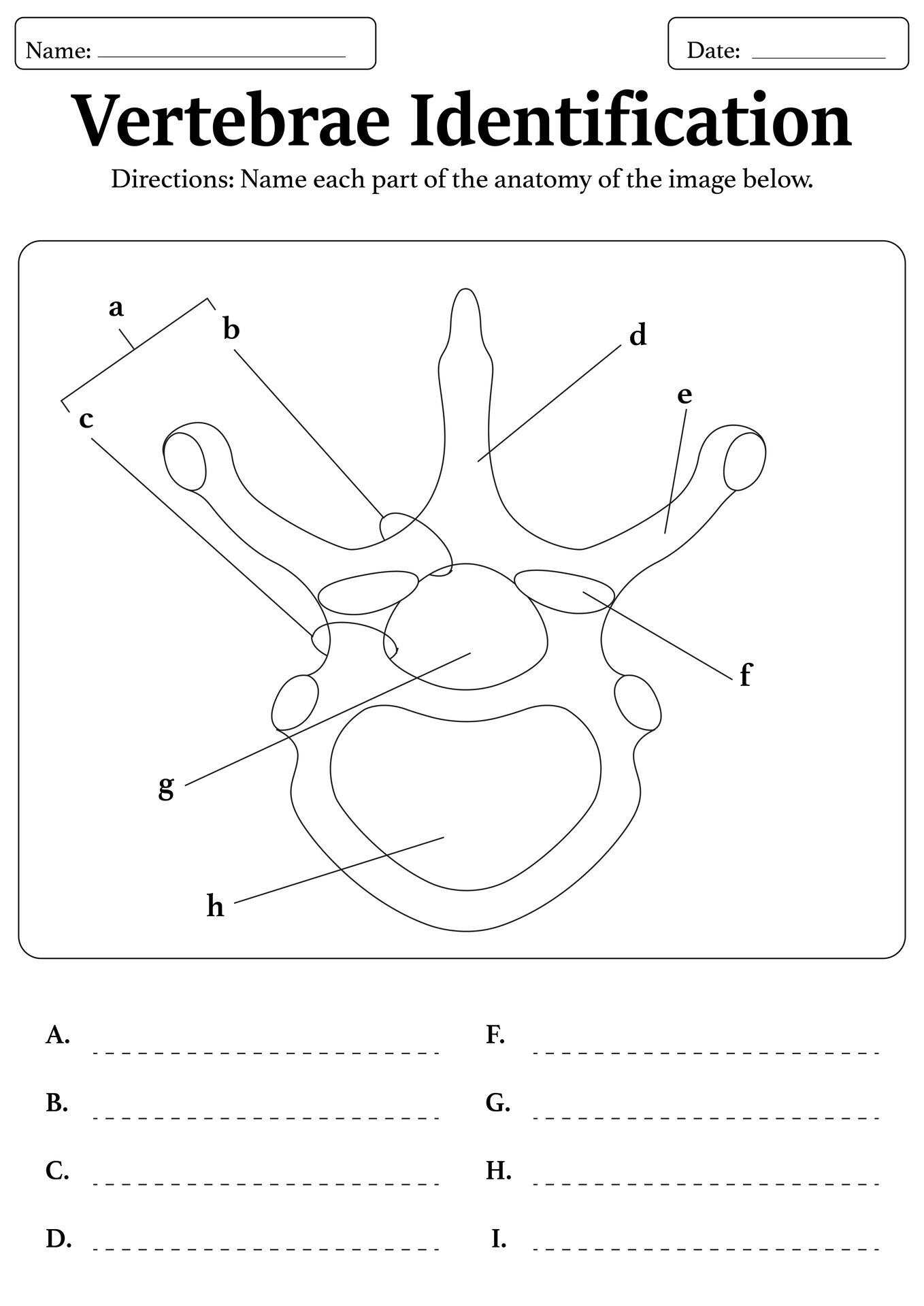
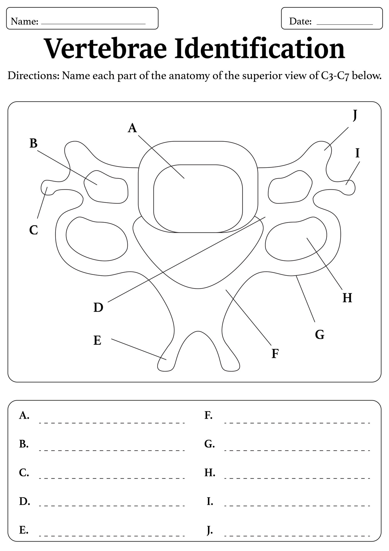
 Vertebrae Identification Labeling Worksheet
 Anatomy Class Vertebrae Labeling Practice
More Other Worksheets
Kindergarten Worksheet My RoomSpanish Verb Worksheets
Spring Clothes Worksheet
Healthy Eating Plate Printable Worksheet
Cooking Vocabulary Worksheet
My Shadow Worksheet
Large Printable Blank Pyramid Worksheet
Relationship Circles Worksheet
DNA Code Worksheet
Meiosis Worksheet Answer Key
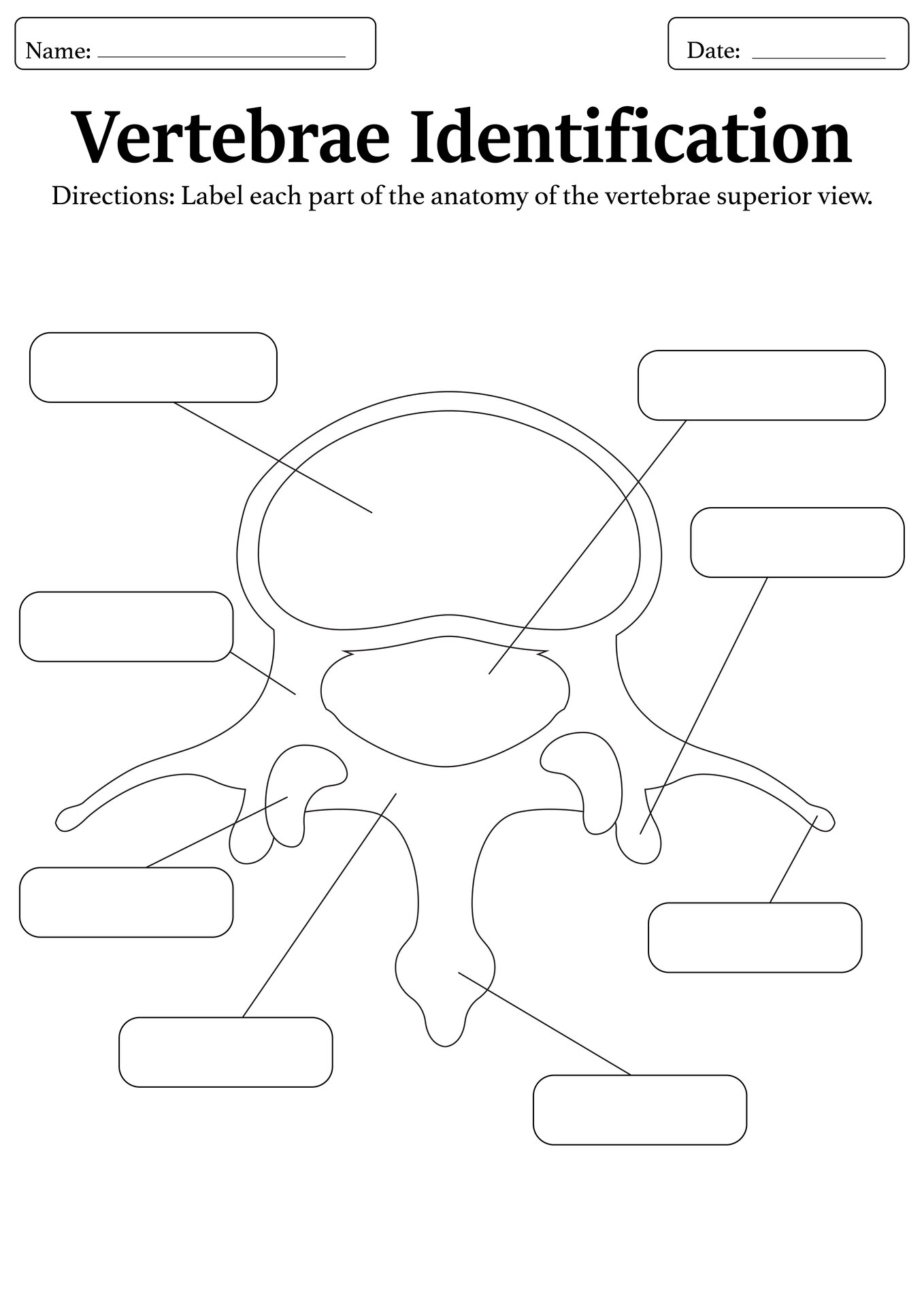
Are you looking for a helpful resource to teach students about the anatomy of vertebrae? Look no further! We have a vertebrae labeling worksheet for you.
This worksheet is a learning tool that allows students to practice identifying and labeling the different parts of the vertebrae. This hands-on activity helps students visualize the structure of the spine and understand the function of each component. By completing a vertebrae worksheet, students can enhance their knowledge of human anatomy in a fun and interactive way.
By incorporating this hands-on activity into your lesson plans, you can enhance students' understanding, engagement, and retention of information. So, use this vertebrae labeling worksheet in your classroom today for an enriching learning experience!
Learn how to effectively incorporate a vertebrae labeling worksheet into your lesson plans!
What is a Vertebrae Labeling Worksheet?
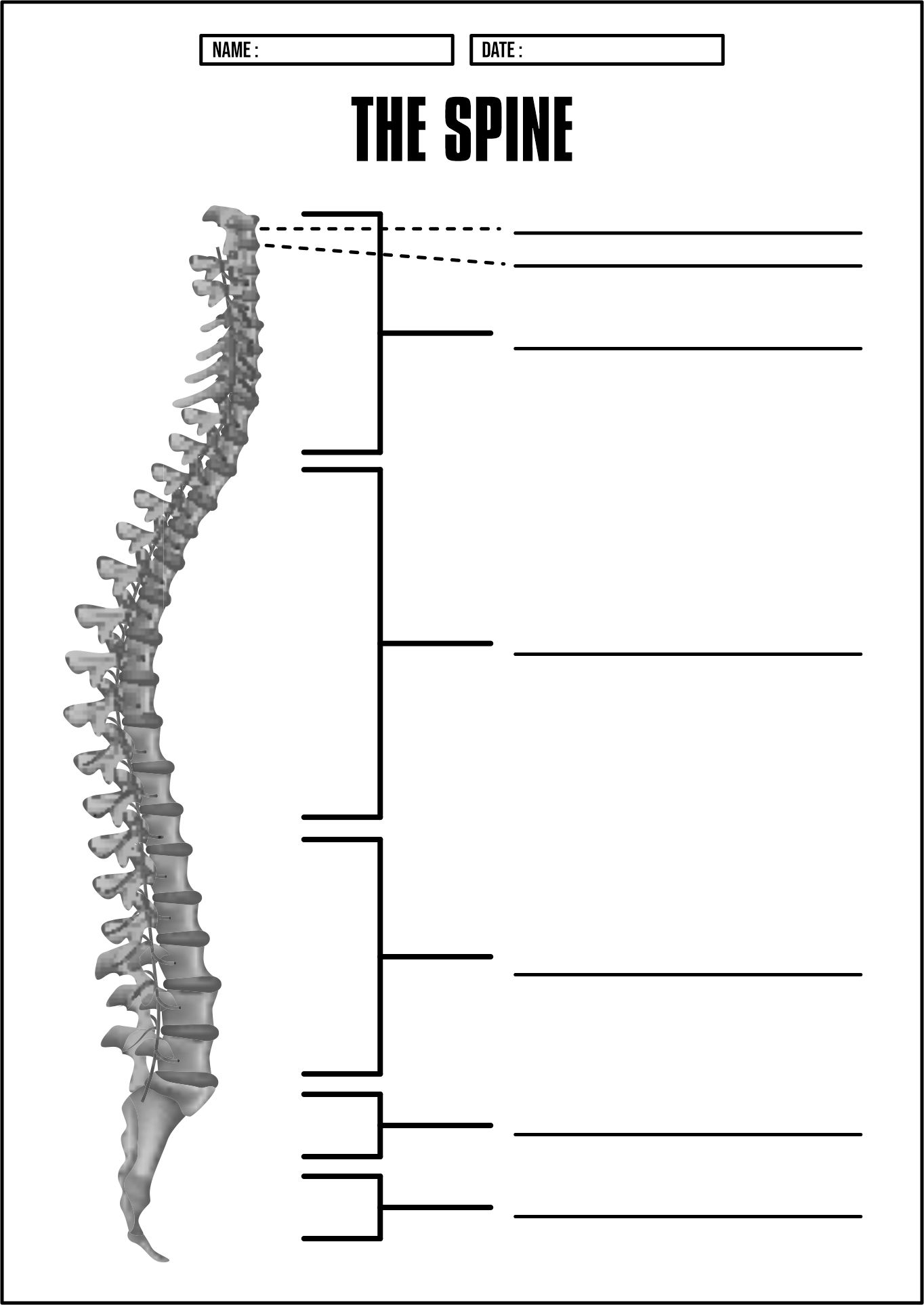
A Vertebrae Labeling Worksheet is an educational tool that is used to help students learn about the different parts of the spine, also known as vertebrae. These worksheets typically include a diagram of the human spine with various labels, such as cervical vertebrae, thoracic vertebrae, lumbar vertebrae, and sacrum. Students are then tasked with labeling each part correctly, helping them to memorize the names and locations of each vertebra.
If you're interested in incorporating these worksheets into your lesson plans, there are many online resources where you can find free printable worksheets like our blog. We offer a variety of anatomy-themed worksheets, including those focused on the vertebrae.
What are the Different Types of Vertebrae Covered in a Vertebrae Labeling Worksheet?
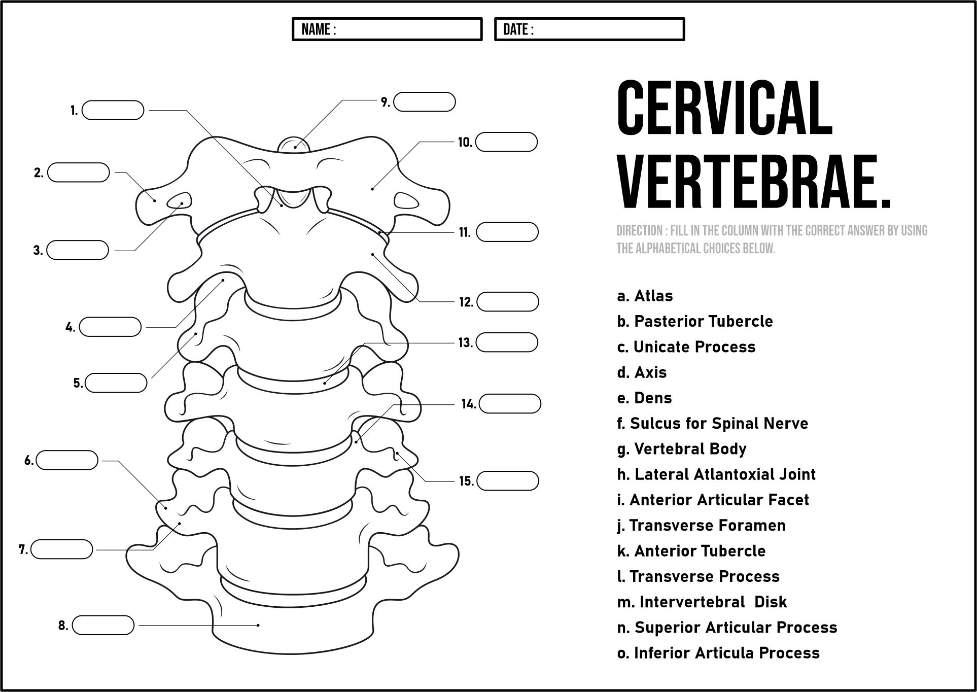
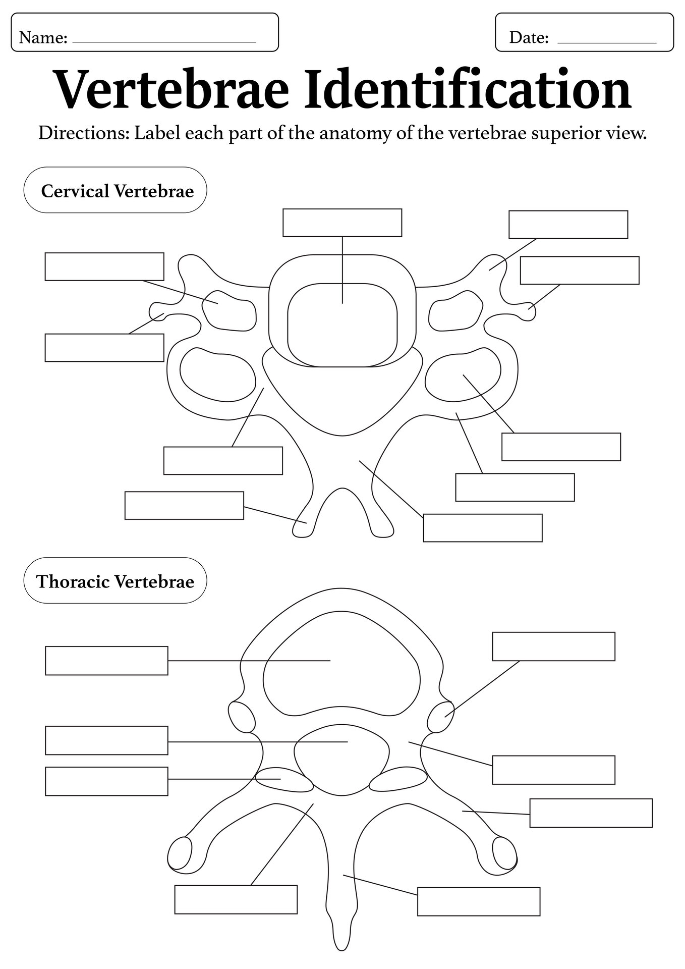
- Cervical Vertebrae: Located in the neck, the cervical vertebrae are the first seven bones of the spinal column. These vertebrae are responsible for supporting the weight of the head and allowing for a wide range of motion in the neck. The cervical vertebrae are labeled C1 through C7, with each vertebra having its own specific characteristics.
- Thoracic Vertebrae: The thoracic vertebrae are the next twelve vertebrae in the spinal column, located in the middle back region. Compared to the cervical vertebrae, these bones are thicker and stronger due to their role in holding up the rib cage. The thoracic vertebrae are labeled T1 through T12, with each vertebra having attachment points for the ribs.
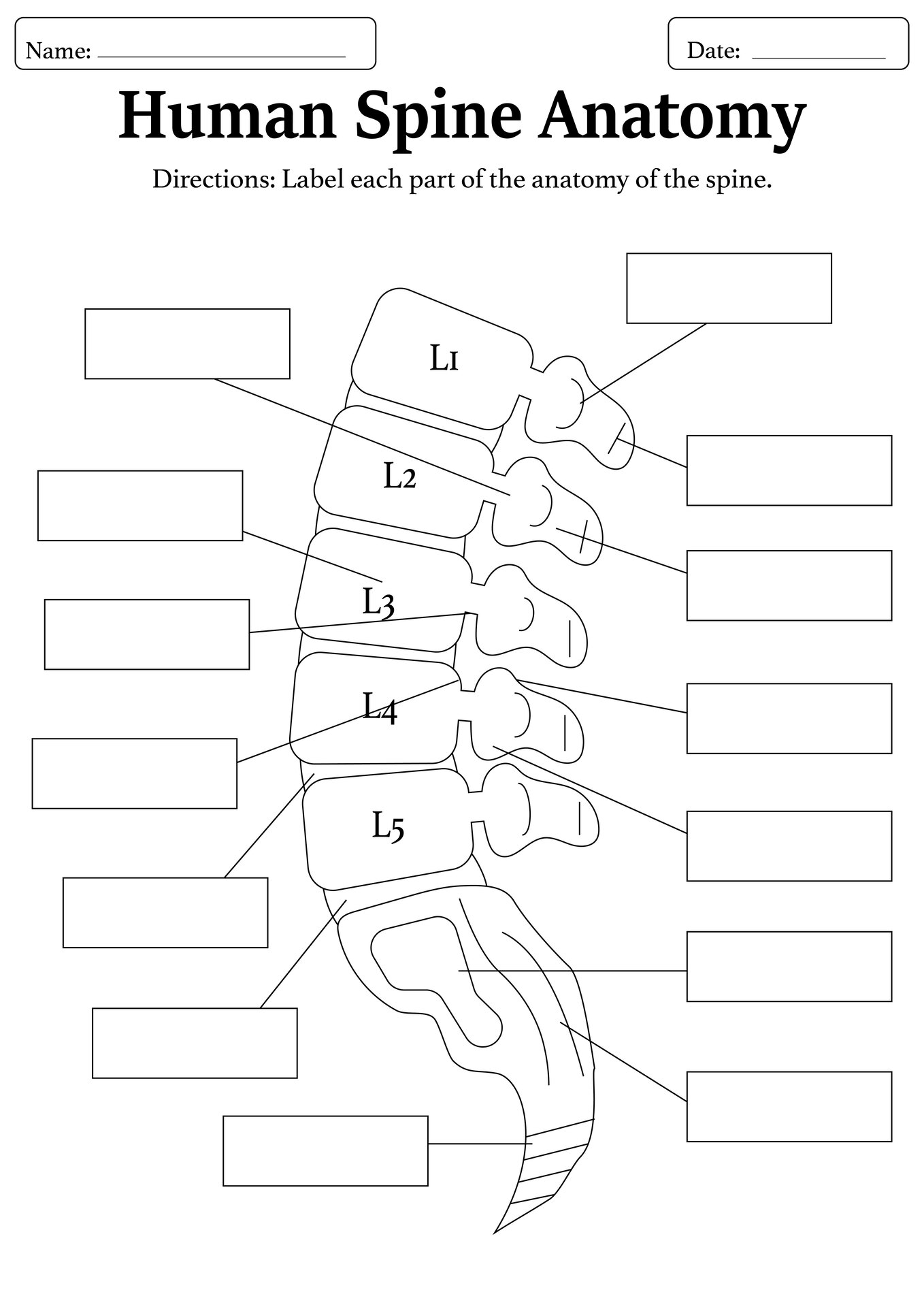
- Lumbar Vertebrae: The lumbar vertebrae are the five largest and strongest vertebrae in the spinal column, located in the lower back region. These vertebrae bear the most weight and are responsible for supporting the majority of the body's weight. The lumbar vertebrae are labeled L1 through L5, with each vertebra being massive in size compared to the cervical and thoracic vertebrae.
- Sacrum: The sacrum is a triangular-shaped bone located at the base of the spine, below the lumbar vertebrae. This part connects the spine to the pelvis and helps support the pelvic area.
- Coccyx: The coccyx, also known as the tailbone, is the small, triangular bone located at the very bottom of the spinal column. The coccyx, made up of four fused vertebrae, acts as a connection point for several muscles, tendons, and ligaments. Although small, it helps support the body when sitting.
Can This Vertebrae Labeling Worksheet Be Used for Both Beginners and Advanced Students?
Yes! Let's find out how to use this worksheet for different skill levels.
- For Beginners: For beginners, a vertebrae worksheet can be an excellent starting point. It provides a basic overview of the spine and its individual vertebrae, allowing students to familiarize themselves with the terminology and layout. With clear labels and simple instructions, beginners can easily identify and learn about the different parts of the spine.
- For Advanced Students: On the other hand, advanced students can benefit from a vertebrae worksheet by delving deeper into the details. They can use the worksheet to test their knowledge and expertise in spinal anatomy, challenging themselves to recall and label each vertebra accurately. This allows advanced students to hone their skills and expand their understanding of the complexities of the spine.
How Can a Vertebrae Labeling Worksheet Be Integrated with Other Anatomy Lessons?
- Incorporate into a Spinal Cord Unit: Begin by introducing the vertebrae worksheet as part of a broader unit on the spinal cord. Encourage students to label the vertebrae while discussing the relationship between the spinal cord and surrounding structures. This hands-on activity can solidify students' understanding of spinal anatomy.
- Connect with Musculoskeletal System Lessons: Integrate the vertebrae worksheet with lessons on the musculoskeletal system to demonstrate how the vertebrae support and protect the spinal cord. Ask students to identify muscle attachment points on the vertebrae, reinforcing the interconnected nature of the musculoskeletal system.
- Cross-Reference with Neuroanatomy Topics: Challenge students to label the vertebrae in relation to key neuroanatomical structures, such as nerve roots and vertebral arteries. By cross-referencing anatomy topics, students can appreciate the functional significance of the vertebrae within the broader context of neuroanatomy.
So, What Can You Learn from a Vertebrae Labeling Worksheet?
- Understanding the Structure of the Spine: One of the main things you can learn from a vertebrae worksheet is the structure of the spine. The spine is made up of several vertebrae that help protect the spinal cord and support the body. By labeling each vertebra on a worksheet, you can visualize the different parts of the spine and how they connect to form a flexible and strong structure.
- Identifying the Different Types of Vertebrae: Another important aspect of using a vertebrae labeling worksheet is the opportunity to identify the different types of vertebrae in the spine. There are five main types of vertebrae: cervical, thoracic, lumbar, sacral, and coccygeal. Each type has unique characteristics and functions, and by labeling them on a worksheet, you can better understand their placement and role in the spine.
- Learning the Importance of Spinal Health: Spinal health is crucial for overall well-being, and a vertebrae worksheet can help you learn more about how to maintain a healthy spine. By labeling each vertebra and understanding its function, you can appreciate the importance of proper posture, regular exercise, and good nutrition in keeping your spine strong and flexible.
In short, vertebrae labeling worksheets are a fantastic tool for anatomy students to reinforce their knowledge of the spine. By utilizing these worksheets effectively and regularly, students can improve their anatomy skills and excel in their studies. So, download a vertebrae worksheet today and take your anatomy learning to the next level!


Have something to share?
Who is Worksheeto?
At Worksheeto, we are committed to delivering an extensive and varied portfolio of superior quality worksheets, designed to address the educational demands of students, educators, and parents.


Comments