Tooth Anatomy Worksheets
Are you a dental student or a science enthusiast looking to expand your knowledge of tooth anatomy? Look no further! In this blog post, we will explore a range of worksheets that are perfect for learning about the different components and functions of teeth. Whether you're studying for an exam or simply wanting to delve deeper into the subject, these worksheets will provide you with a valuable learning tool.
Table of Images 👆
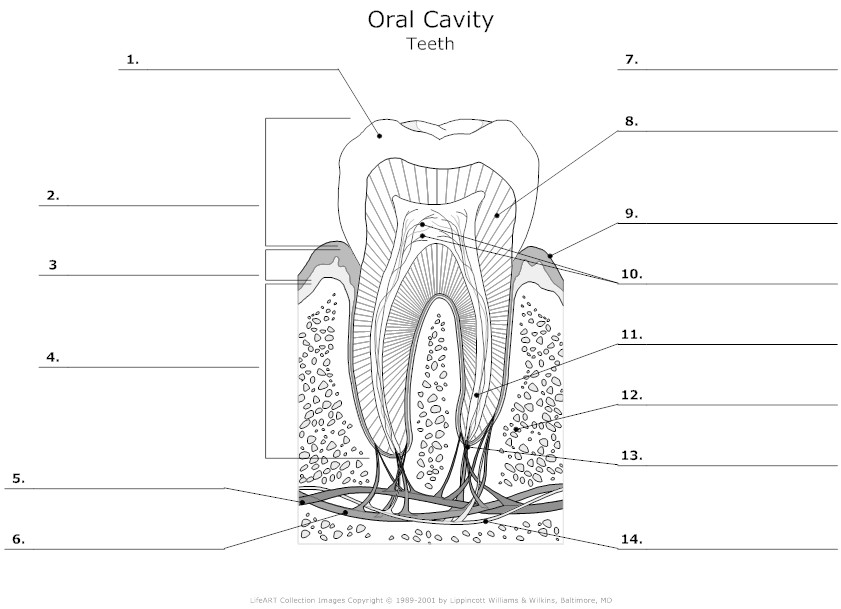
 Tooth Anatomy Diagram Unlabeled

 Teeth Diagram Worksheet
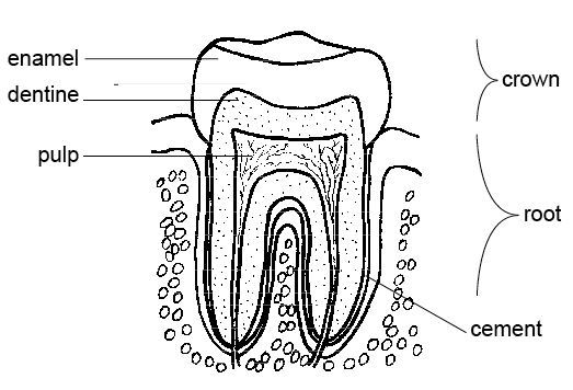
 Anatomy Tooth Coloring Page
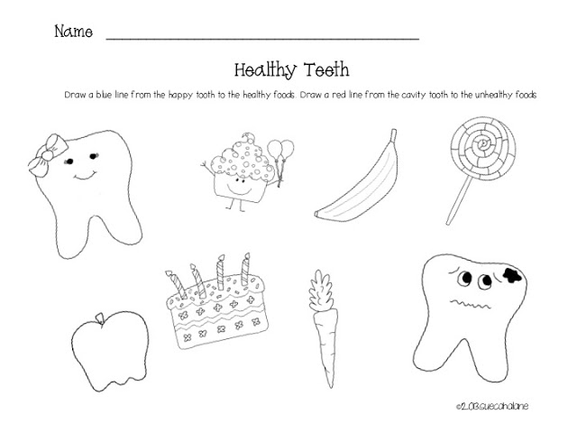
 Healthy Teeth Worksheet
 Printable Tooth Anatomy
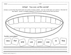
 High Frequency Words Worksheets
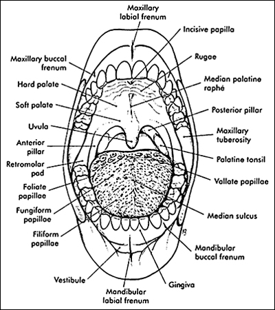
 Mouth Diagram Labeled
More Other Worksheets
Kindergarten Worksheet My RoomSpanish Verb Worksheets
Cooking Vocabulary Worksheet
DNA Code Worksheet
Meiosis Worksheet Answer Key
Art Handouts and Worksheets
7 Elements of Art Worksheets
All Amendment Worksheet
Symmetry Art Worksheets
Daily Meal Planning Worksheet
What is the crown of a tooth?
The crown of a tooth is the visible, white part of the tooth that is above the gumline. It is the part of the tooth that is used for chewing and grinding food.
What is the function of the enamel?
Enamel is the hard, outer layer of the tooth that serves to protect the tooth from wear and decay. It is the strongest substance in the human body and acts as a barrier against bacterial invasion and damage from acidic foods and drinks. Enamel also helps to insulate the tooth against temperature changes and provides a smooth surface for chewing and grinding food.
What is the dentin and what role does it play?
Dentin is a hard tissue that makes up the bulk of a tooth, located beneath the enamel layer. It serves as a protective layer that surrounds the inner pulp of the tooth, providing structural support and insulation. Dentin also helps to protect the more sensitive pulp and nerve tissues from external stimuli like temperature changes or trauma. Additionally, it is responsible for transmitting sensations such as pressure or touch to the nerves in the pulp.
What is the pulp chamber and what does it contain?
The pulp chamber is the innermost part of a tooth that houses the dental pulp, which consists of nerves, blood vessels, and connective tissue. This pulp plays a crucial role in the tooth's development and health, as it provides nourishment and helps detect sensation and pain.
What are the different types of teeth in the human mouth?
In the human mouth, there are four main types of teeth: incisors, canines, premolars, and molars. Incisors are used for cutting and chopping food, canines are for tearing and grasping, premolars help with chewing and grinding, and molars are also used for chewing and grinding. Each type of tooth has a specific function in the process of breaking down food for digestion.
What are the roots of a tooth and how are they attached?
The roots of a tooth are the part of the tooth that extends below the gumline and anchor the tooth in the jawbone. They are attached to the jawbone through a specialized connective tissue called the periodontal ligament, which securely holds the tooth in place while allowing for some flexibility to absorb the forces of chewing and biting. The roots also contain blood vessels and nerves that supply nutrients to the tooth and enable sensation.
What is the periodontal ligament and what is its function?
The periodontal ligament is a group of connective tissue fibers that attaches the tooth to the surrounding alveolar bone within the jaw. Its main function is to support the tooth by acting as a cushion during biting and chewing, thus providing stability and flexibility while also helping to sense the forces applied to the tooth. Additionally, the periodontal ligament plays a crucial role in maintaining dental health by aiding in the remodeling of the surrounding bone in response to the stresses placed on the tooth.
What is the cementum and where is it located?
Cementum is a hard tissue found on the outer surface of a tooth's root. It serves as a protective layer and helps anchor the tooth to the surrounding bone through the periodontal ligament. The cementum is essential for maintaining the stability and health of the tooth within the jawbone.
What are the different parts of a tooth's anatomy?
The different parts of a tooth's anatomy include the crown (the visible part above the gum line), the root (the part below the gum line that anchors the tooth in place), the enamel (the hard outer layer protecting the tooth), the dentin (inner layer beneath the enamel), the pulp (soft tissue at the center of the tooth containing nerves and blood vessels), and the gum line (where the tooth meets the gum tissue).
What is the significance of understanding tooth anatomy for dental health?
Understanding tooth anatomy is crucial for dental health as it allows for recognizing potential issues early on. By knowing the different parts of a tooth, such as enamel, dentin, pulp, and roots, individuals can better comprehend how diseases like decay or infection can impact specific areas and seek appropriate treatment. Additionally, understanding tooth anatomy helps in practicing good oral hygiene habits, as knowledge of tooth structure can aid in effective brushing and flossing techniques to prevent plaque buildup and maintain overall oral health.


Have something to share?
Who is Worksheeto?
At Worksheeto, we are committed to delivering an extensive and varied portfolio of superior quality worksheets, designed to address the educational demands of students, educators, and parents.


Comments