Microscope Lab Worksheet
If you're a curious student or an enthusiastic science teacher searching for a comprehensive and helpful resource to enhance your microscope lab experience, then this blog post is for you. Whether you're exploring the fascinating world of cells or observing intricate microorganisms, a well-designed microscope lab worksheet can provide guidance and structure to ensure a successful and meaningful laboratory session.
Table of Images 👆
- Compound Microscope Lab Activity
- Label Microscope Parts Worksheet
- Microscope Lab Sheet for Kids
- Microscope Parts Worksheet Answers
- Microscope Lab Worksheet Answers
- Letter E Microscope Lab Activity
- Compound Microscope Worksheet
- Onion Cell Wet Mount Microscope Slide
- Cell Microscope Lab Worksheet
- E Microscope Lab Worksheet
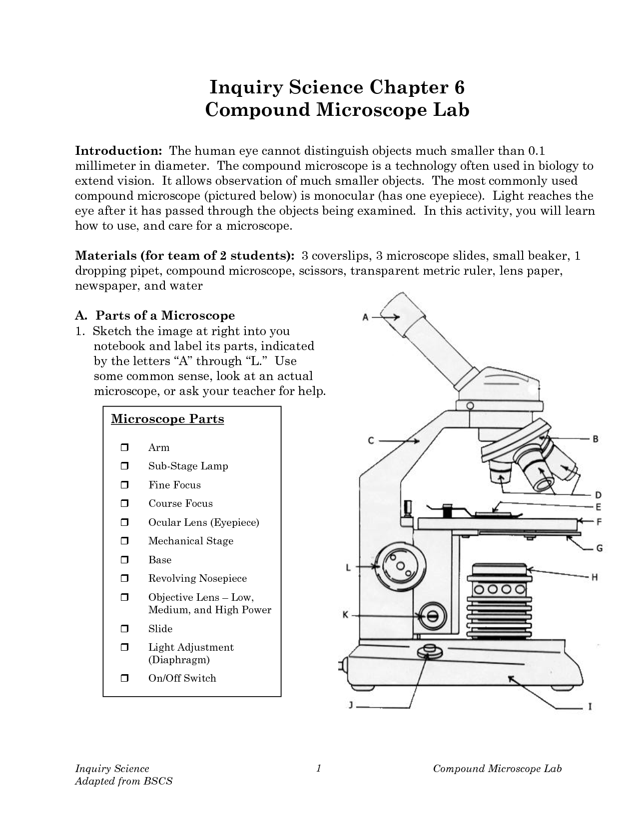
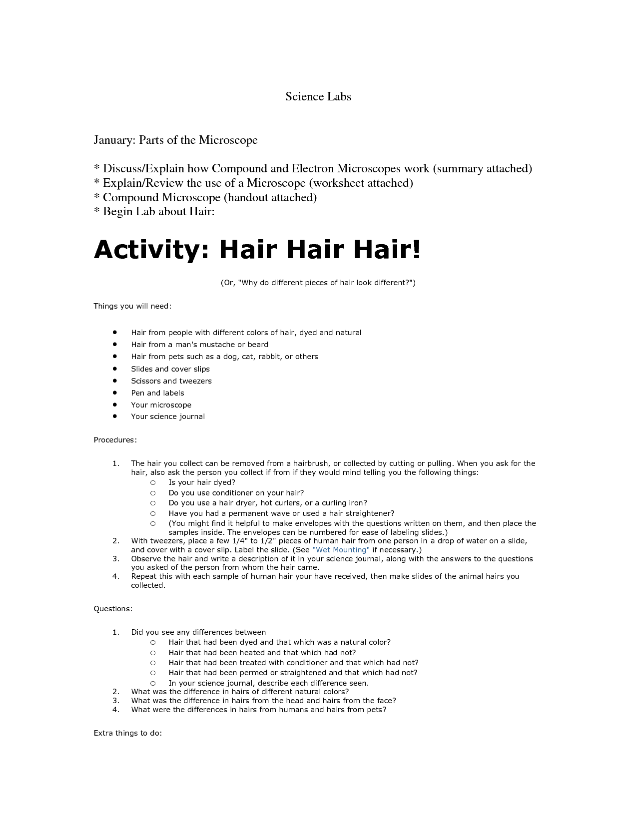
 Compound Microscope Lab Activity
 Compound Microscope Lab Activity
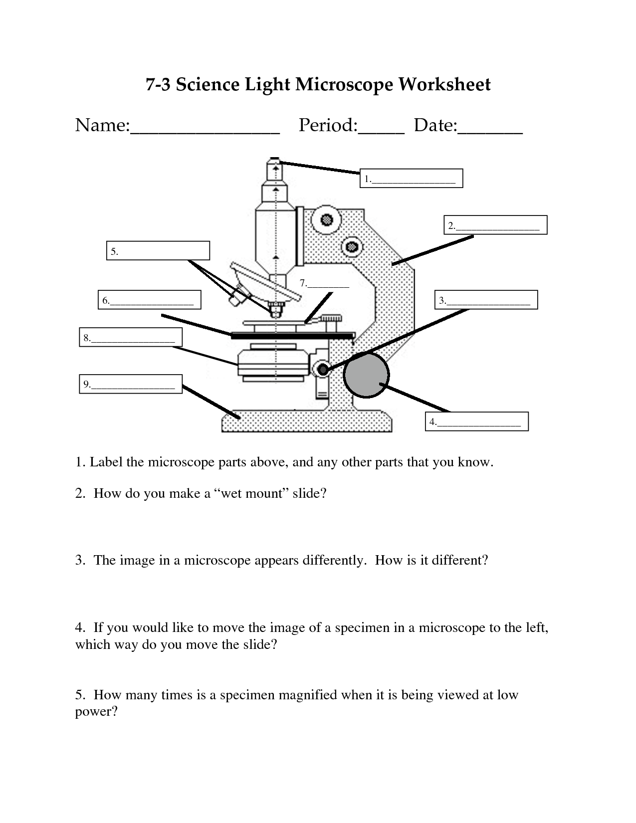
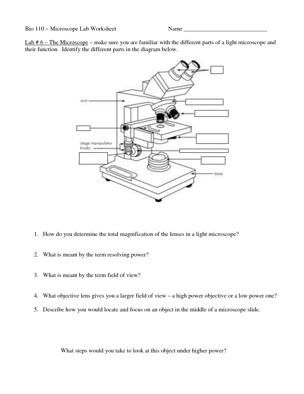
 Label Microscope Parts Worksheet
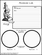
 Microscope Lab Sheet for Kids
 Microscope Parts Worksheet Answers
 Microscope Lab Worksheet Answers
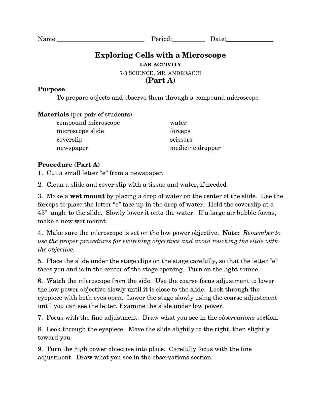
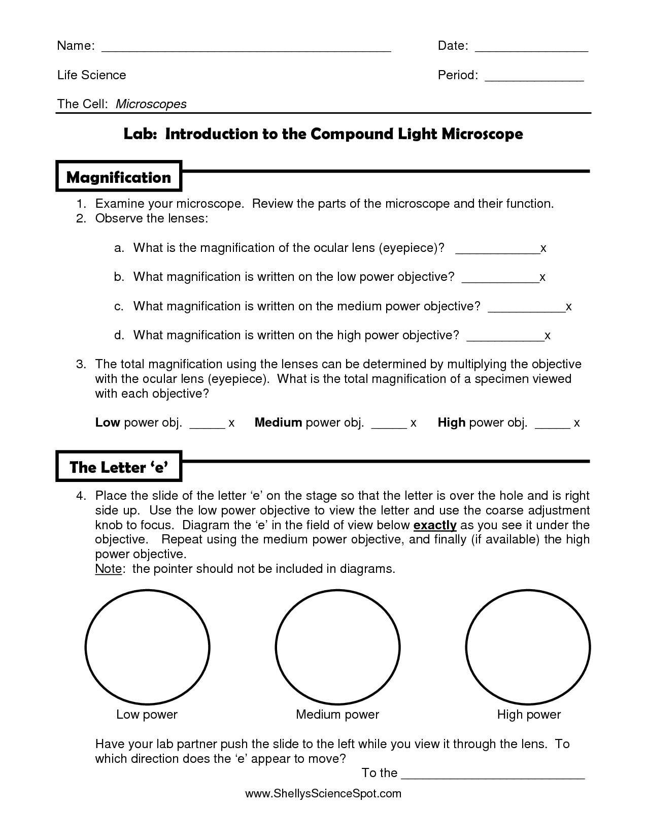
 Letter E Microscope Lab Activity
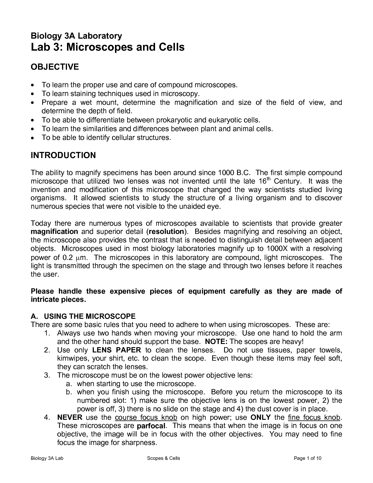
 Compound Microscope Worksheet
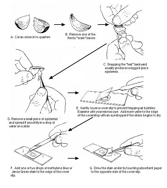
 Onion Cell Wet Mount Microscope Slide
 Cell Microscope Lab Worksheet
 E Microscope Lab Worksheet
 Letter E Microscope Lab Activity
More Other Worksheets
Kindergarten Worksheet My RoomSpanish Verb Worksheets
Cooking Vocabulary Worksheet
DNA Code Worksheet
Meiosis Worksheet Answer Key
Art Handouts and Worksheets
7 Elements of Art Worksheets
All Amendment Worksheet
Symmetry Art Worksheets
Daily Meal Planning Worksheet
What is the purpose of using a microscope in a lab?
The purpose of using a microscope in a lab is to magnify and visualize objects that are too small to be seen with the naked eye. This tool enables scientists to study the structure, morphology, and characteristics of microscopic organisms, cells, tissues, or particles in detail, aiding in scientific research, medical diagnostics, and various other laboratory processes.
What is the difference between magnification and resolution in microscopy?
Magnification in microscopy refers to the ability to enlarge the image of a sample to see details that may not be visible to the naked eye, whereas resolution refers to the ability to distinguish fine details and separate two distinct points or objects in an image. In other words, magnification increases the size of the image, while resolution determines the level of clarity and sharpness in the details of that image.
How do you calculate total magnification on a compound microscope?
To calculate the total magnification on a compound microscope, multiply the magnification of the objective lens by the magnification of the eyepiece lens. For example, if the objective lens has a magnification of 40x and the eyepiece lens has a magnification of 10x, the total magnification would be 40x * 10x = 400x.
What is the role of the objective lens in a microscope?
The objective lens in a microscope is responsible for magnifying the specimen that is being observed. It is the primary lens that focuses the light onto the specimen and produces an enlarged image. The objective lens often has varying magnification levels, allowing users to switch between different levels of magnification.
How does adjusting the fine focus knob affect the image in a microscope?
Adjusting the fine focus knob in a microscope allows for precise focusing of the image by making small changes in the distance between the objective lens and the specimen. This helps to bring different layers of the specimen into sharp focus and enhances the clarity and resolution of the image being viewed under the microscope.
What are the steps for properly cleaning and storing a microscope after use?
After using a microscope, the first step is to turn it off and unplug it. Remove any slides and clean the lenses using lens paper or a soft cloth with lens cleaning solution. Next, clean the outside of the microscope with a mild disinfectant wipe. Carefully store the microscope in a clean and dry cabinet, preferably covered to prevent dust accumulation. Make sure to properly secure any accessories such as the eyepieces and objectives. Always follow the manufacturer's instructions for specific cleaning and maintenance guidelines to ensure the longevity and performance of the microscope.
What are the main components of a compound microscope?
The main components of a compound microscope include the eyepiece lens, objective lens, stage, stage clips, focus knobs, light source, and base. The eyepiece lens is where the viewer looks through, while the objective lens magnifies the specimen. The stage is where the specimen is placed and held in position by stage clips. Focus knobs are used to adjust the focus of the specimen. The light source illuminates the specimen, and the base provides support and stability for the microscope.
Why is it important to start with the lowest magnification objective when using a microscope?
It is important to start with the lowest magnification objective when using a microscope because it provides a wider field of view and a greater depth of field, making it easier to locate and focus on the specimen. Starting with the lowest magnification also helps prevent the risk of damaging the microscope or specimen by accidentally hitting the lens or slide while trying to adjust the focus at higher magnifications. Additionally, adjusting the focus and centering the specimen at a lower magnification can improve the overall quality of the image when switching to higher magnifications.
What is the function of the diaphragm or iris aperture in a microscope?
The diaphragm or iris aperture in a microscope regulates the amount of light passing through the specimen and into the lenses, allowing the user to control the brightness and contrast of the image being viewed. By adjusting the size of the aperture, the user can improve the clarity and visibility of the specimen under observation.
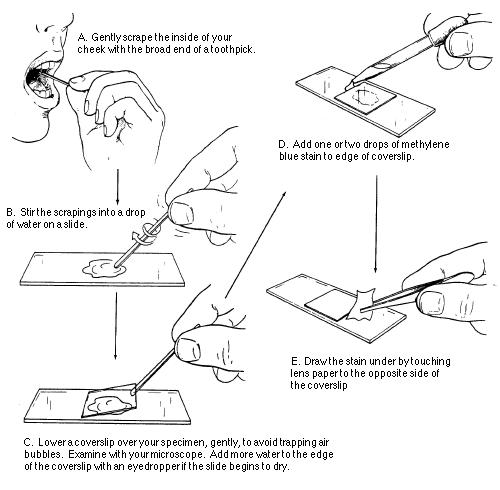
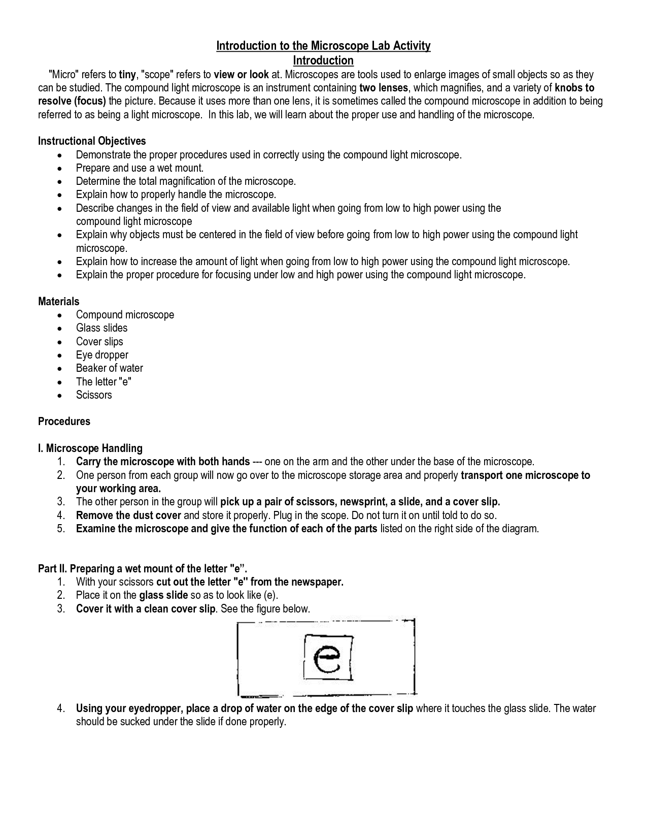
How can you ensure proper slide preparation for viewing under a microscope?
To ensure proper slide preparation for viewing under a microscope, start by cleaning the microscope slide and cover slip thoroughly to remove any dirt or debris. Then, place a small drop of the specimen you want to view in the center of the slide. Gently lower a cover slip onto the drop of specimen to avoid trapping air bubbles. Secure the cover slip in place and make sure there are no gaps between the slide and cover slip. Finally, label the slide with the specimen's name or relevant information and store it in a slide box to prevent damage or contamination before viewing under a microscope.


Have something to share?
Who is Worksheeto?
At Worksheeto, we are committed to delivering an extensive and varied portfolio of superior quality worksheets, designed to address the educational demands of students, educators, and parents.


Comments