Microscope Diagram Worksheet
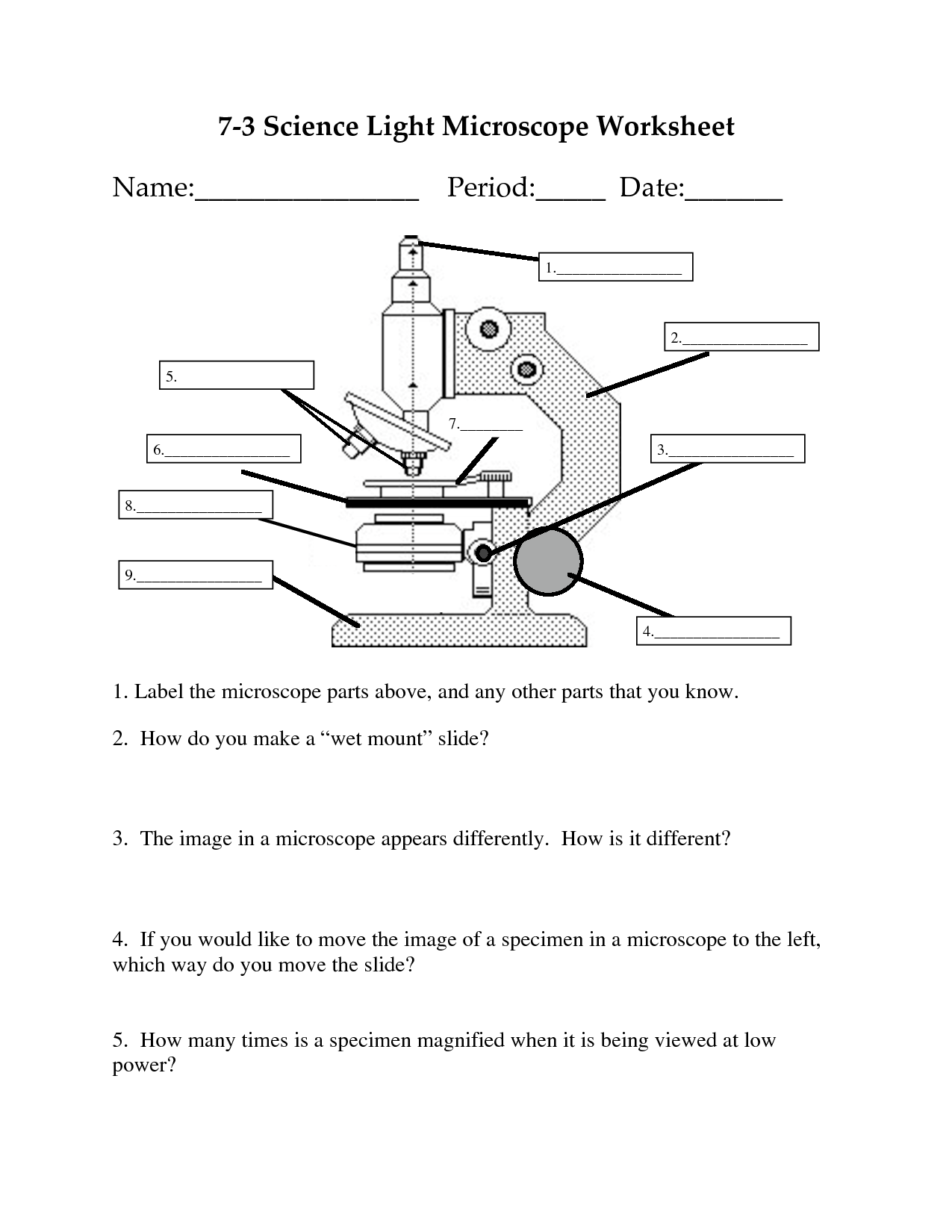
A microscope diagram worksheet is a helpful tool for students studying biology or any science-related subjects that involve the use of microscopes. This worksheet provides a detailed illustration of a microscope and its different parts, allowing students to practice labeling each component and understand their functions better. Whether you're a teacher looking for a resource to reinforce the concept of microscopy or a student wanting to test your knowledge, a microscope diagram worksheet is a valuable entity for enhancing your understanding in this subject.
Table of Images 👆
- Compound Light Microscope Parts Worksheet
- Compound Light Microscope Parts Blank
- Microscope Parts Worksheet
- Fill in the Blank Microscope Diagram
- Microscope with Labeled Parts
- Label Microscope Parts Worksheet
- Animal Cell Coloring Worksheet Answers
- Scanning Electron Microscope Diagram
- Compound Microscope Clip Art
- Printable Book Report Worksheet
- Evolution Worksheet Answer Key
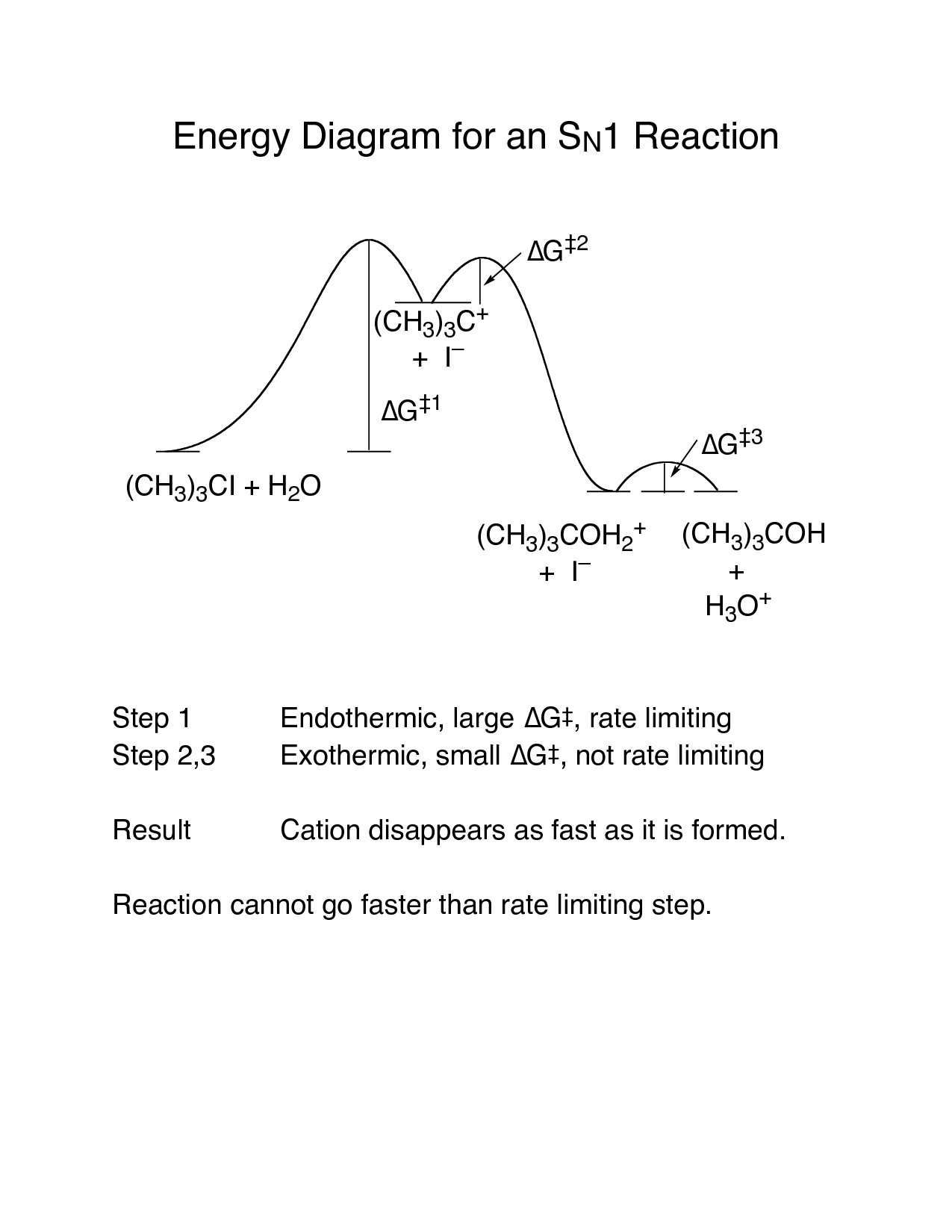
- SN1 Energy Reaction Diagram
- Mass and Weight Measurement Conversion Chart
More Other Worksheets
Kindergarten Worksheet My RoomSpanish Verb Worksheets
Cooking Vocabulary Worksheet
DNA Code Worksheet
Meiosis Worksheet Answer Key
Art Handouts and Worksheets
7 Elements of Art Worksheets
All Amendment Worksheet
Symmetry Art Worksheets
Daily Meal Planning Worksheet
What is the purpose of a microscope diagram worksheet?
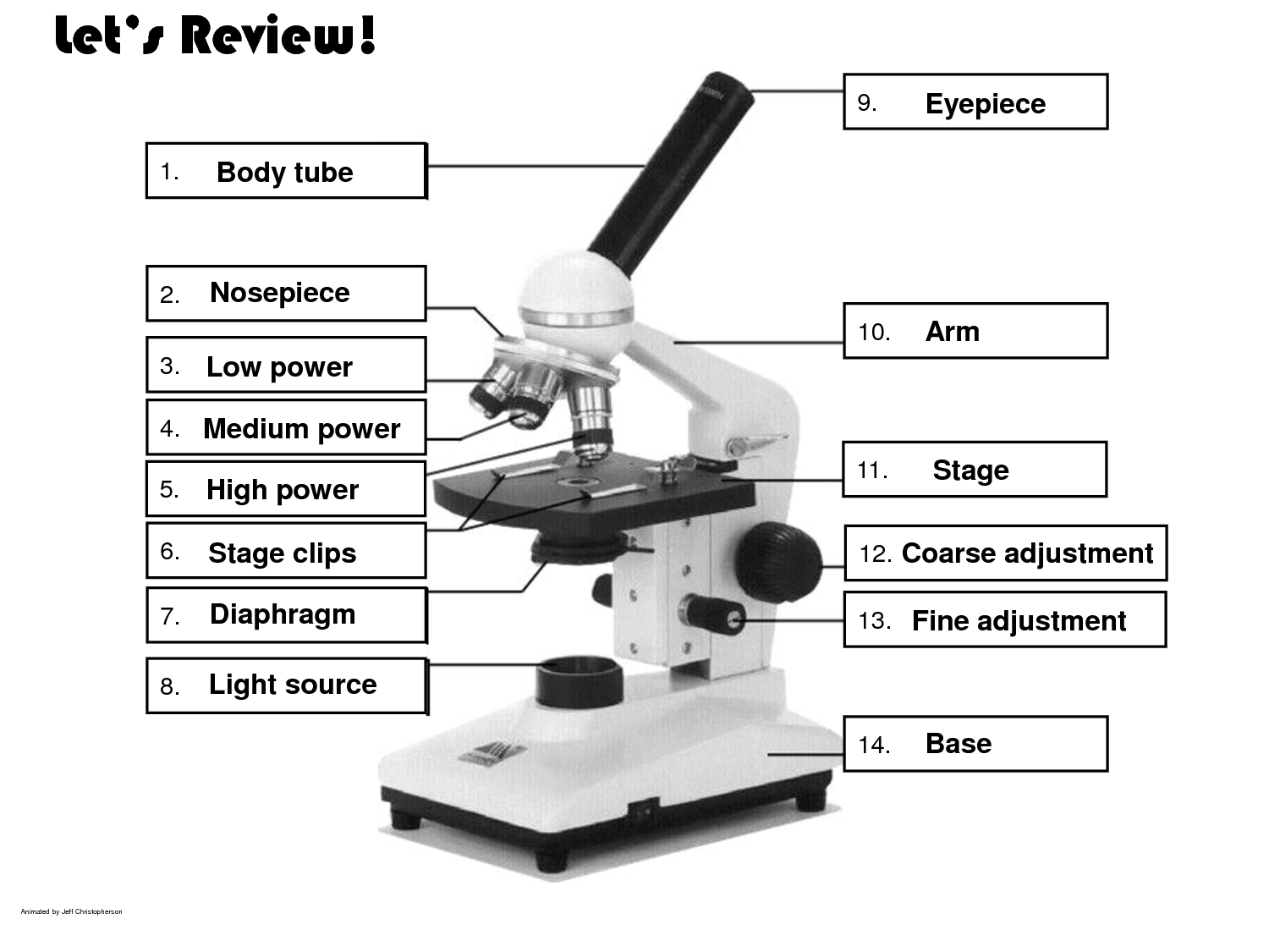
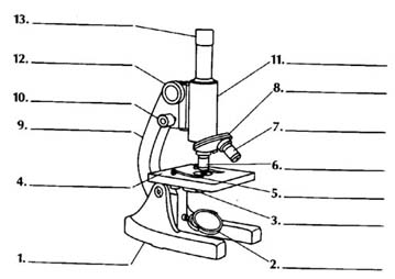
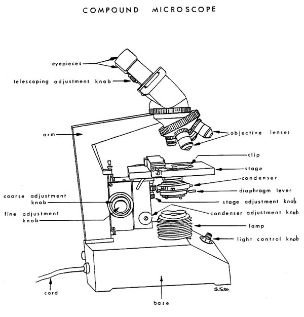
A microscope diagram worksheet serves the purpose of helping students learn and understand the parts and functions of a microscope. It allows students to visually explore and label the different components of a microscope, such as the eyepiece, objective lenses, stage, and diaphragm, reinforcing their knowledge of how microscopes work and how to use them effectively for scientific observation and experimentation.
What are the different parts of a microscope that are typically labeled in the diagram?
The different parts of a microscope that are typically labeled in a diagram include the eyepiece, objective lens, stage, focus knobs, diaphragm, light source, and arm.
Why is it important to label the parts of a microscope correctly?
It is important to label the parts of a microscope correctly to ensure accurate communication and understanding among users. Properly labeled parts help users identify and locate specific components, follow operating instructions effectively, and troubleshoot any issues that may arise during microscope use. This can lead to improved efficiency, consistency, and precision in scientific experiments, research, and education involving microscopes.
How do the eyepiece and objective lenses work together to magnify the specimen?
The eyepiece and objective lenses work together in a compound microscope to magnify the specimen. The objective lens collects light and forms a magnified real image of the specimen, which is then further magnified by the eyepiece. The eyepiece acts as a magnifying glass to enlarge the image produced by the objective lens, allowing the user to see the specimen at a higher level of magnification. By combining the magnification powers of both lenses, the compound microscope is able to provide a more detailed view of the specimen.
What is the function of the stage on a microscope?
The stage on a microscope serves as a platform to hold the specimen being observed. It allows the specimen to be easily positioned and moved so that different areas can be examined under the lenses of the microscope. The stage also often includes scales or markings for measuring the size or distance of the specimen being viewed.
Explain the role of the diaphragm in controlling the amount of light that passes through the specimen.
The diaphragm in a microscope controls the amount of light that reaches the specimen by adjusting the size of the opening in the condenser. By changing the size of the aperture, the user can regulate the intensity and focus of the light that illuminates the specimen, ultimately affecting the clarity and contrast of the image produced. This feature allows for better control over the lighting conditions and enhances the quality of the viewing experience when observing microscopic specimens.
What is the purpose of the coarse and fine adjustment knobs on a microscope?
The purpose of the coarse adjustment knob on a microscope is to make large changes in focus quickly by moving the stage closer to or further away from the objective lens, while the fine adjustment knob is used for making small, precise focus adjustments to bring the specimen into sharp focus. The coarse adjustment is for initial focusing, and the fine adjustment is for fine-tuning the focus for clear and detailed observation.
Why is it important to use a coverslip when examining a specimen under the microscope?
Using a coverslip when examining a specimen under the microscope is important because it flattens the specimen, prevents it from drying out, and minimizes the risk of damaging the high-power objective lens. It also helps in enhancing the clarity and resolution of the image by reducing the amount of light refraction and distortion that can occur when viewing the specimen directly without a coverslip.
How does the condenser lens help improve the quality of the image?
The condenser lens helps improve the quality of the image by focusing and directing light rays onto the specimen being observed. This ensures that the specimen is evenly illuminated and enhances contrast, making details clearer and more visible. Additionally, the condenser lens helps to increase the resolution of the image by reducing scatter and improving the overall sharpness of the specimen under the microscope.
What are some safety precautions that should be taken when using a microscope?
When using a microscope, always ensure that you handle the equipment with care to prevent damage or injury. Be sure to keep the area well-lit and clean to avoid accidents or contamination. Additionally, remember to always use lens paper to clean the lenses gently and avoid scratching them. Remember to secure slides properly on the stage to prevent them from falling, and always start with the lowest magnification to avoid damaging the slides or lenses. Lastly, when adjusting the focus, move the stage slowly to prevent sudden movements that could damage the equipment or the specimen on the slide.
Have something to share?
Who is Worksheeto?
At Worksheeto, we are committed to delivering an extensive and varied portfolio of superior quality worksheets, designed to address the educational demands of students, educators, and parents.









Comments