Leg Anatomy Worksheets
Are you eager to master the intricate details of leg anatomy? Look no further than our comprehensive leg anatomy worksheets. These educational resources are designed to help students, medical professionals, and anatomy enthusiasts deepen their understanding of the structure and function of the leg.
Table of Images 👆
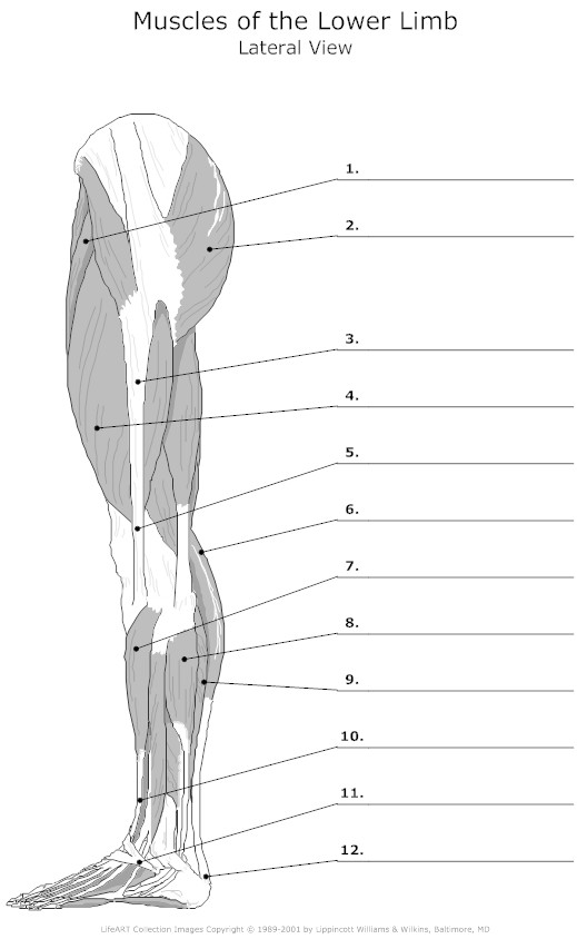
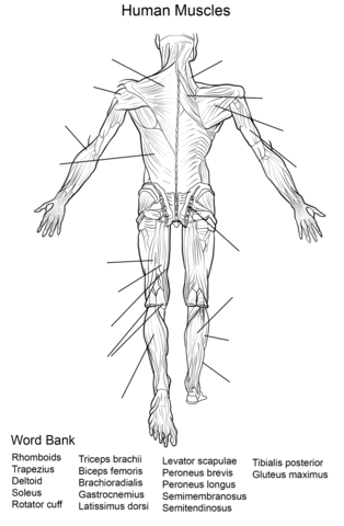
- Lower Limb Muscles Anatomy Worksheet
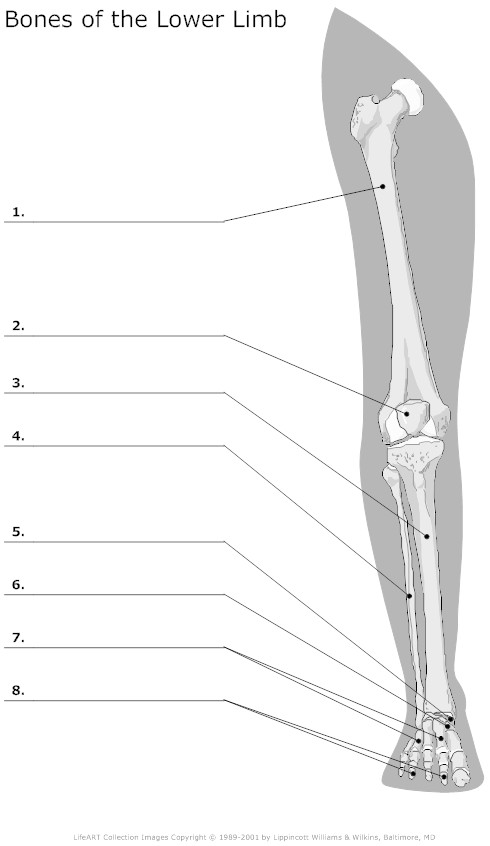
- Lower Limb Bones Unlabeled
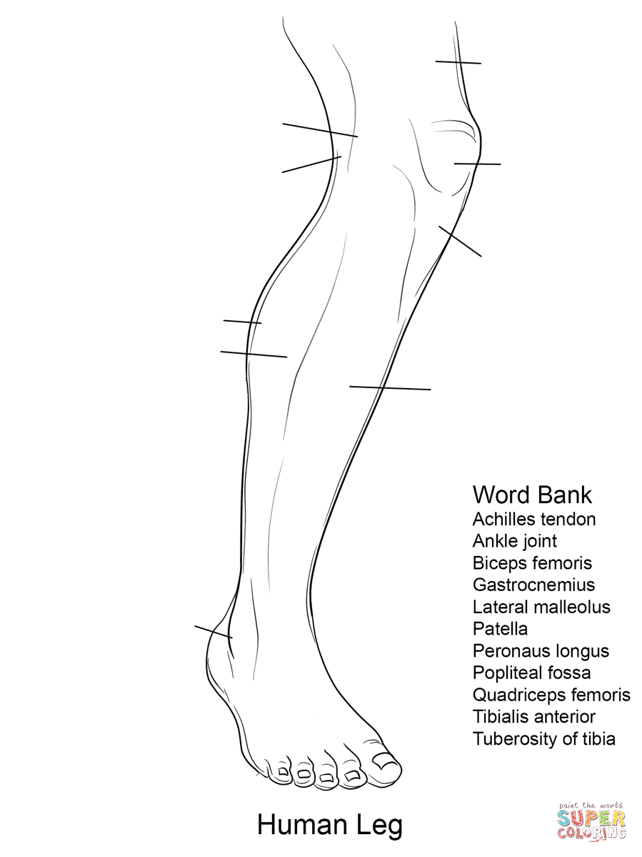
- Free Printable Human Anatomy Coloring Pages
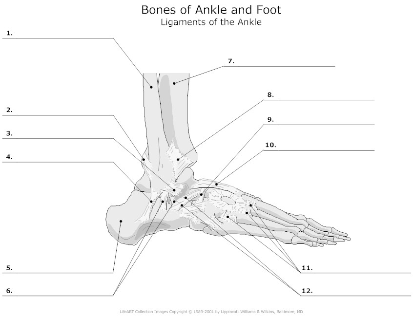
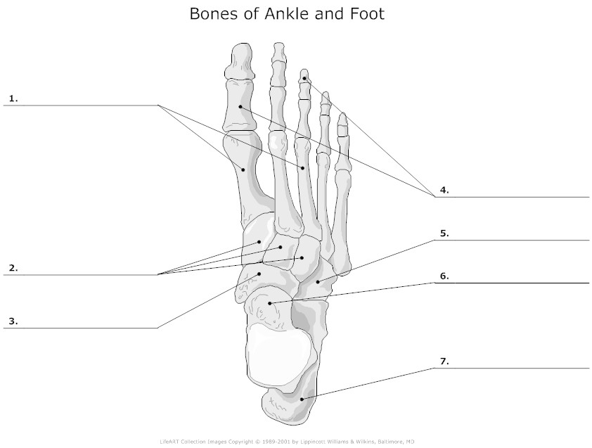
- Foot and Ankle Bones Unlabeled
- Lower Limb Blood Vessel Coloring Page
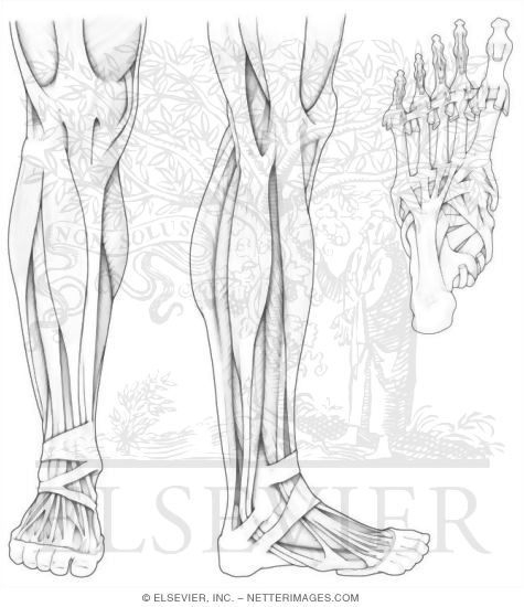
- Lower Leg Muscle Diagram Blank
- Anatomy Muscle Worksheets Printable
- Human Anatomy Coloring Pages Printable
- Lower Leg Muscle Labeling Blank Worksheet
- Foot Bone Diagram Unlabeled
- Muscle Anatomy Coloring Book Page
- Anatomy Bones Coloring Pages
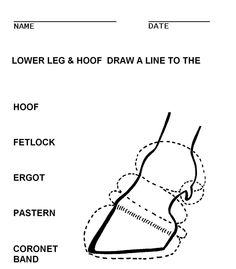
- Horse Saddle Parts Worksheet
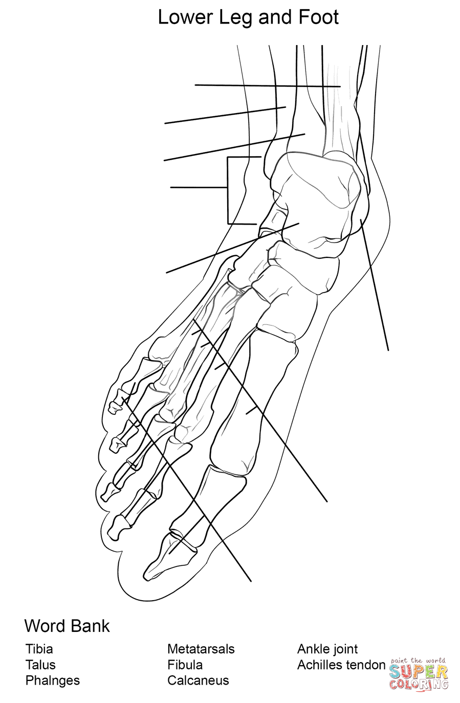
- Lower Leg Bones Diagram
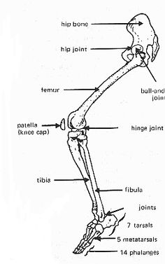
- Diagram of Leg Bones and Joints
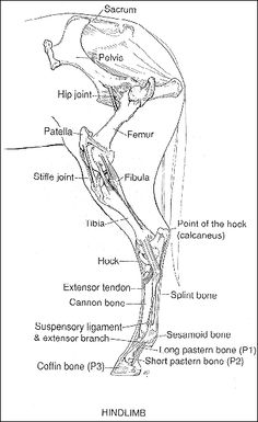
- Horse Stifle Joint Anatomy
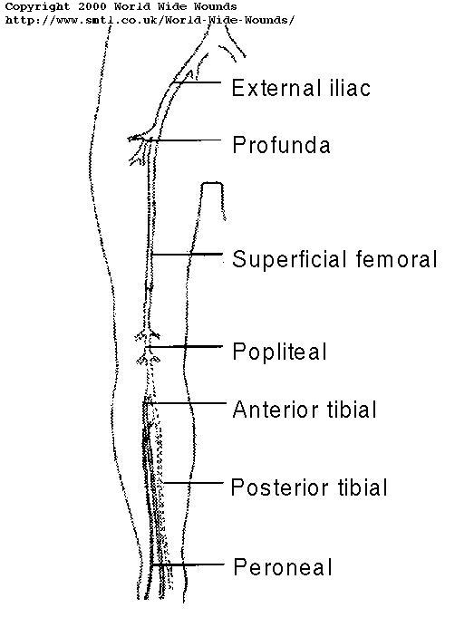
- Lower Extremity Arterial Duplex Worksheet
- Human Anatomy Muscles Coloring Pages Printable
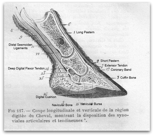
- Horse Anatomy Diagrams Legs
More Other Worksheets
Kindergarten Worksheet My RoomSpanish Verb Worksheets
Cooking Vocabulary Worksheet
DNA Code Worksheet
Meiosis Worksheet Answer Key
Art Handouts and Worksheets
7 Elements of Art Worksheets
All Amendment Worksheet
Symmetry Art Worksheets
Daily Meal Planning Worksheet
What are the major bones of the leg?
The major bones of the leg are the femur (thigh bone), tibia (shin bone), and fibula (calf bone). The femur is the longest and strongest bone in the body, connecting the hip to the knee. The tibia is larger and supports most of the body's weight, while the fibula is located on the outside of the lower leg and helps stabilize the ankle.
What is the function of the patella?
The patella, also known as the kneecap, functions to protect the knee joint and improve the leverage of major thigh muscles when straightening the leg. It acts as a fulcrum, allowing the quadriceps muscles to generate more force for movements such as running, jumping, and kicking. The patella also helps distribute the forces acting on the knee joint more evenly, reducing the risk of injuries and providing stability during various activities.
What are the primary muscles in the anterior compartment of the leg?
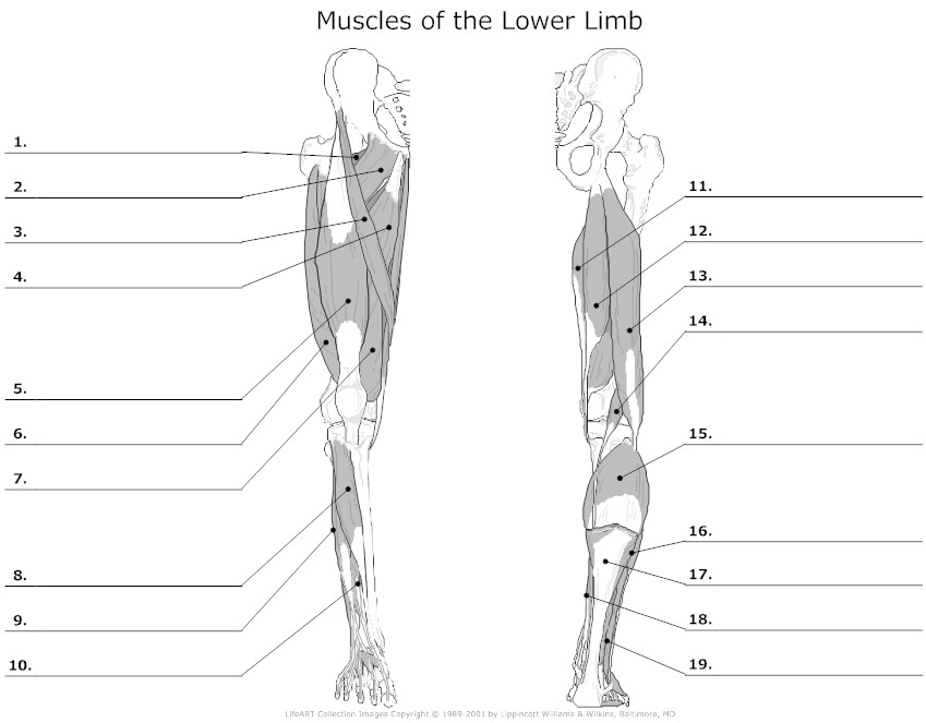
The primary muscles in the anterior compartment of the leg are the tibialis anterior, extensor hallucis longus, extensor digitorum longus, and peroneus tertius. These muscles work together to dorsiflex the foot and extend the toes, as well as aid in inversion and eversion of the foot.
Describe the structure and function of the Achilles tendon.
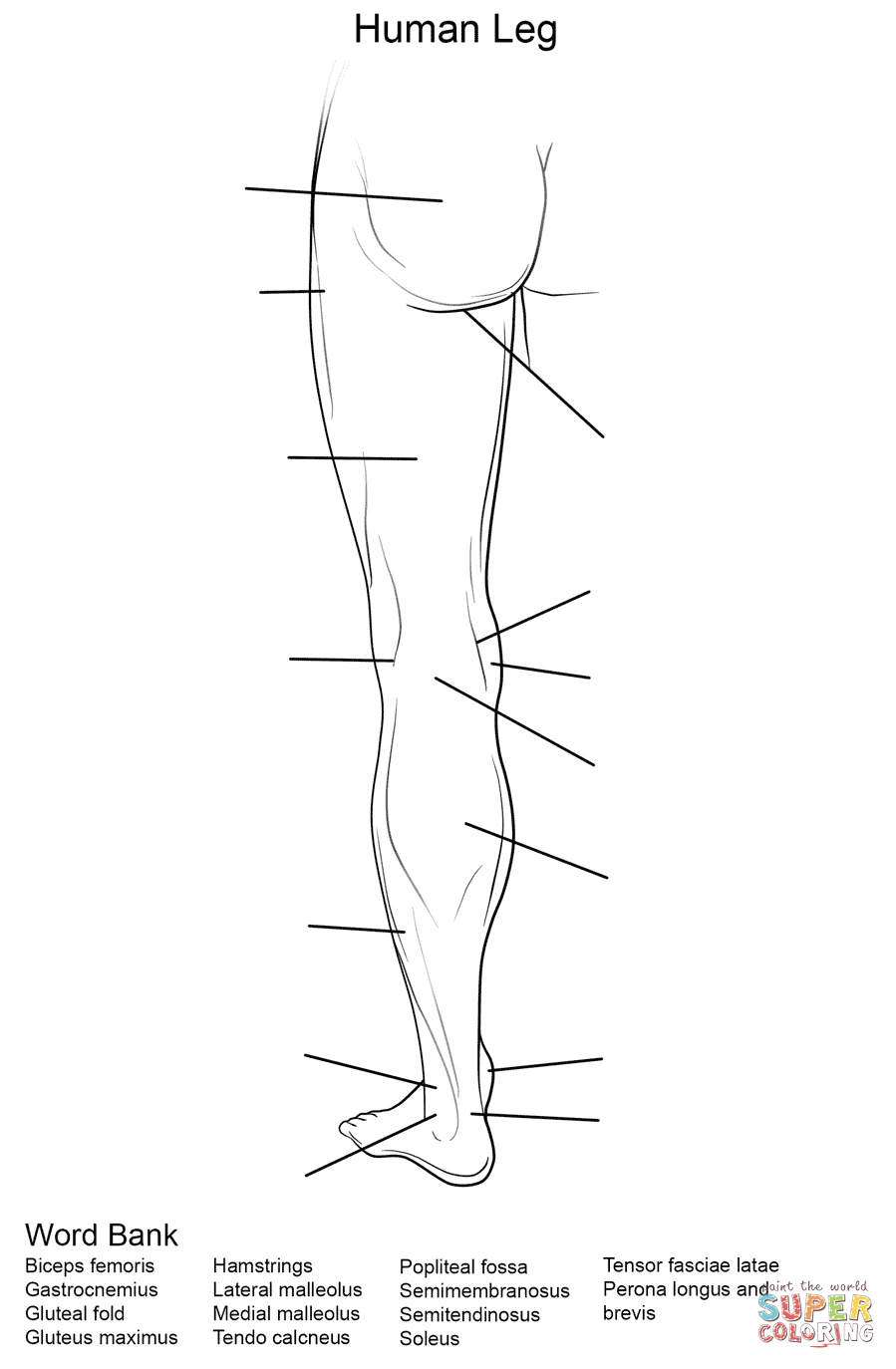
The Achilles tendon is the largest and strongest tendon in the human body, connecting the calf muscles to the heel bone (calcaneus). This fibrous band of tissue enables the contraction of the calf muscles, allowing extension of the foot downward, which is crucial for activities like walking, running, and jumping. The Achilles tendon absorbs the forces generated during these movements, providing stability and power for the lower leg. Additionally, it acts as a spring-like mechanism, storing and releasing energy to enhance performance.
What are the major blood vessels in the leg?
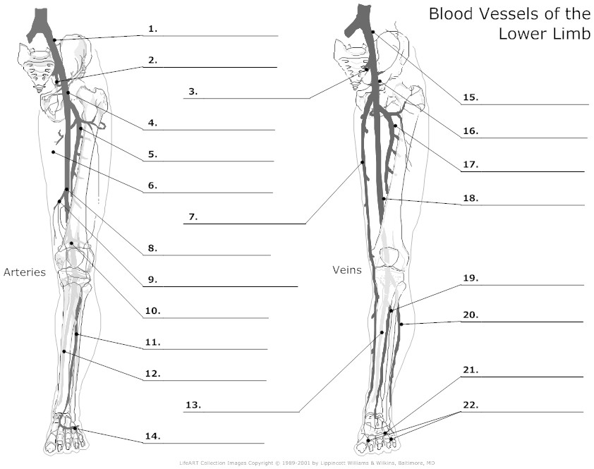
The major blood vessels in the leg are the femoral artery and femoral vein, which run from the hip to the knee and supply blood to the thigh and lower leg. The popliteal artery and popliteal vein are located behind the knee and provide blood flow to the lower leg and foot. Additionally, the anterior and posterior tibial arteries carry blood down the shin and calf, while the dorsalis pedis artery supplies blood to the foot. These blood vessels play a crucial role in maintaining circulation and providing nutrients and oxygen to the tissues in the leg.
Explain the role of ligaments in the stability of the knee joint.
Ligaments play a crucial role in providing stability to the knee joint by connecting the bones and ensuring proper alignment during movement. In the knee joint, the main ligaments are the anterior cruciate ligament (ACL), posterior cruciate ligament (PCL), medial collateral ligament (MCL), and lateral collateral ligament (LCL). These ligaments help to control the range of motion of the knee and prevent excessive movement in directions that could cause injury. The ACL and PCL specifically provide stability by preventing forward and backward sliding of the tibia on the femur, while the MCL and LCL help to stabilize the knee from side-to-side movements. Maintaining the integrity of these ligaments is crucial for the overall stability and function of the knee joint.
What are the main nerves that innervate the leg?
The main nerves that innervate the leg are the femoral nerve, sciatic nerve, tibial nerve, and common fibular (peroneal) nerve. These nerves control the movement and sensation in the thigh, knee, calf, and foot, playing a crucial role in the motor and sensory functions of the leg.
Discuss the difference between the tibia and fibula.
The tibia and fibula are two bones in the lower leg. The tibia, also known as the shinbone, is the larger and stronger of the two bones and is located on the medial (inner) side of the leg. It bears most of the body's weight and is crucial for stability and weight-bearing. The fibula is the thinner bone and is located on the lateral (outer) side of the leg. It plays a supporting role in the leg and is responsible for muscle attachments, but it does not bear as much weight as the tibia. Overall, while both bones are important for the structure and function of the lower leg, the tibia is more prominent in weight-bearing and support, while the fibula plays a secondary role in these functions.
What are the main components of the ankle joint?
The main components of the ankle joint are the tibia, fibula, and talus bones, along with ligaments that provide stability and facilitate movement. The tibia and fibula form the top and sides of the ankle joint, while the talus bone sits below, forming a hinge-like structure that allows for dorsiflexion and plantarflexion movements. The ligaments, including the deltoid and lateral ligaments, help to hold the bones together and provide support during weight-bearing activities.
Describe the movements and range of motion of the hip joint.
The hip joint is a ball-and-socket joint that allows for a wide range of motion. It can flex, extend, abduct (move away from the midline), adduct (move towards the midline), and rotate internally and externally. The hip can move in all planes, making it one of the most versatile weight-bearing joints in the body. Its movements are crucial for walking, running, jumping, and performing various daily activities.
Have something to share?
Who is Worksheeto?
At Worksheeto, we are committed to delivering an extensive and varied portfolio of superior quality worksheets, designed to address the educational demands of students, educators, and parents.











Comments