The Compound Microscope Worksheet Answers
If you're a student or educator looking for a helpful resource to enhance your understanding of compound microscopes, you've come to the right place. In this blog post, we will provide you with a collection of accurate and comprehensive answers to the compound microscope worksheet.
Table of Images 👆
- Compound Light Microscope Parts Worksheet
- Microscope Parts Quiz Worksheet
- Microscope Parts Worksheet
- Label Microscope Parts Worksheet
- Microscope Parts Labeled
- Microscope Diagram Worksheet
- Compound Microscope Worksheet
- Microscope Parts Worksheet Answers
- Cell Microscope Lab Worksheet
- Light Microscope Parts Worksheet
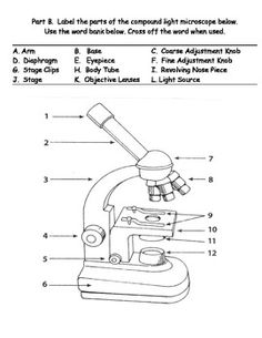
- Microscope Parts Blank Diagram

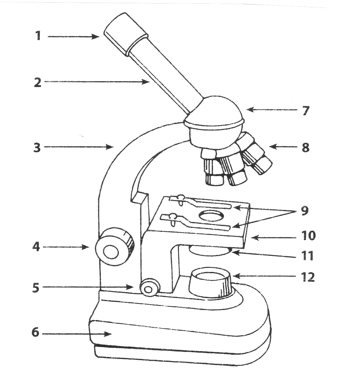
 Compound Light Microscope Parts Worksheet
 Microscope Parts Quiz Worksheet
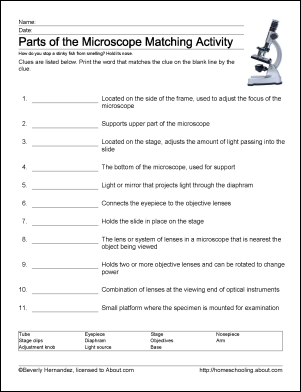
 Microscope Parts Worksheet
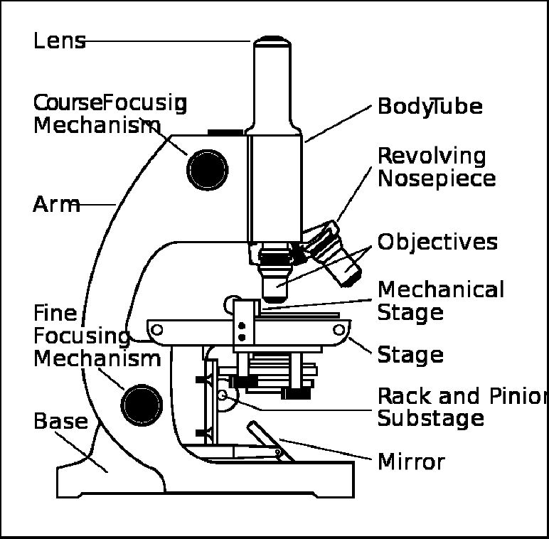
 Label Microscope Parts Worksheet
 Microscope Parts Labeled
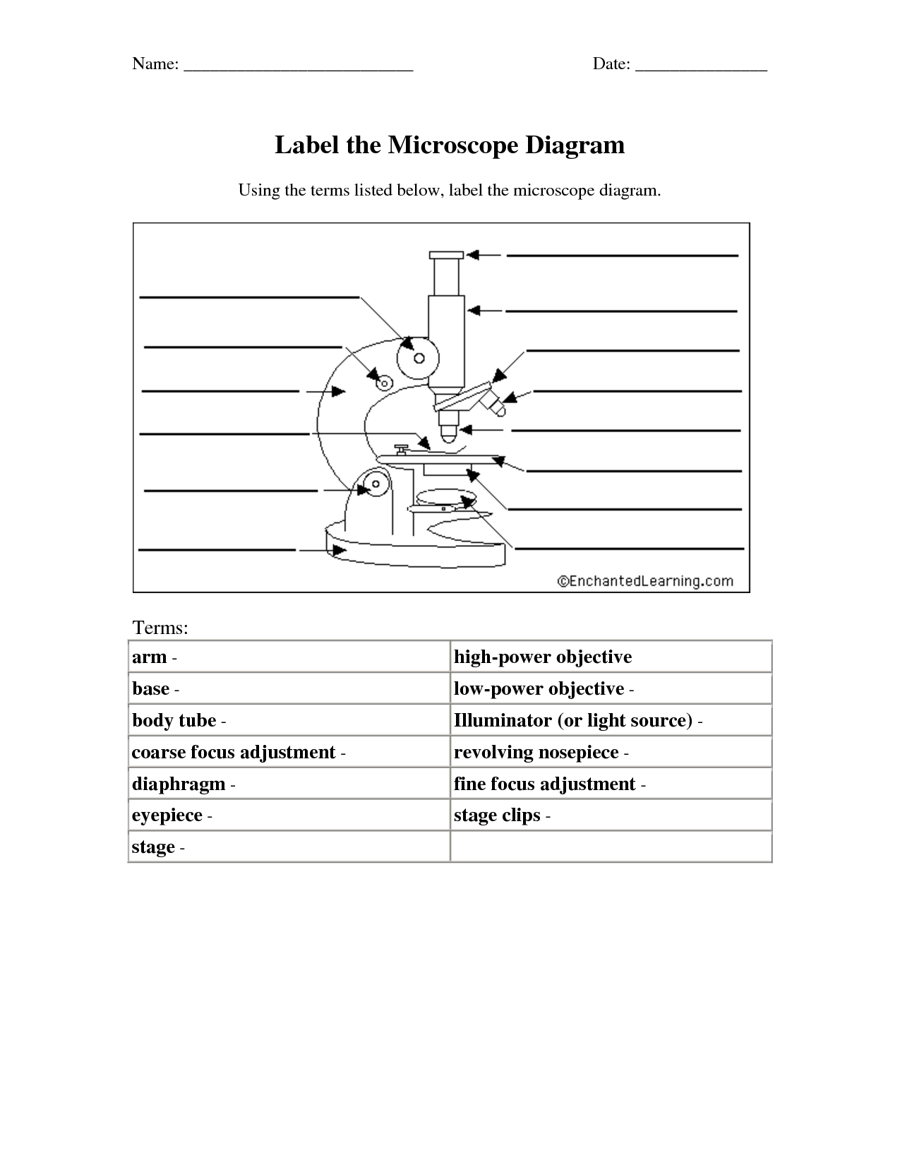
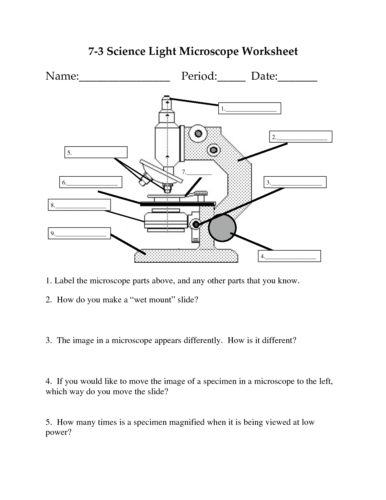
 Microscope Diagram Worksheet
 Microscope Parts Worksheet
 Compound Microscope Worksheet
 Microscope Parts Worksheet Answers
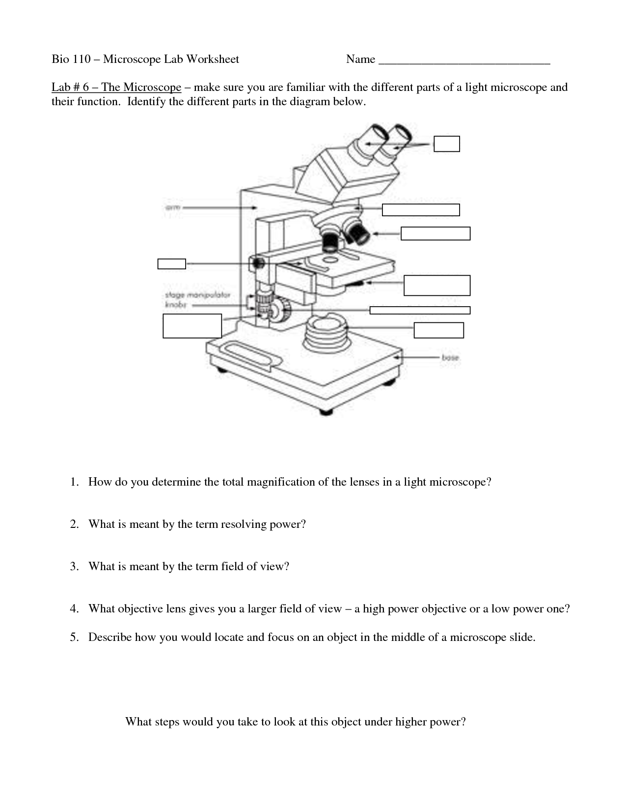
 Cell Microscope Lab Worksheet
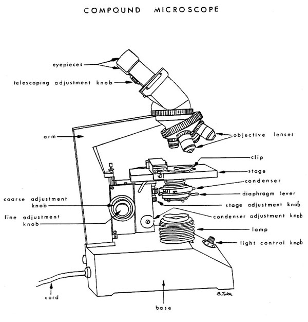
 Light Microscope Parts Worksheet

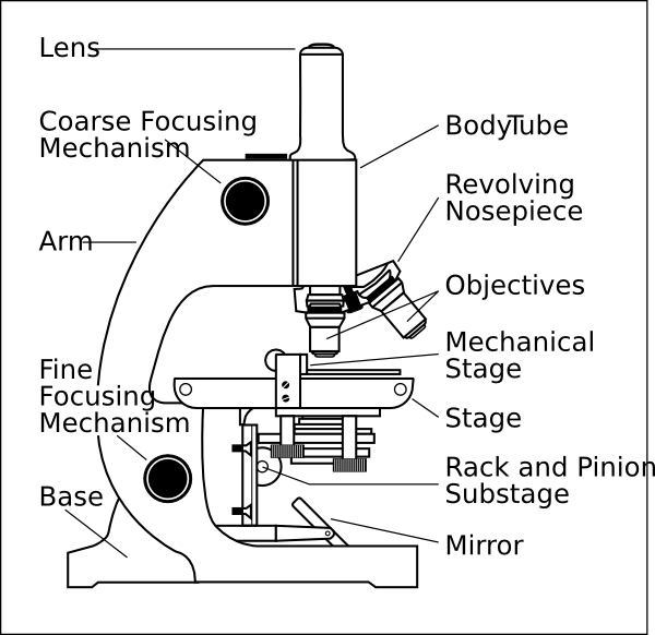
 Microscope Parts Blank Diagram
More Other Worksheets
Kindergarten Worksheet My RoomSpanish Verb Worksheets
Cooking Vocabulary Worksheet
DNA Code Worksheet
Meiosis Worksheet Answer Key
Art Handouts and Worksheets
7 Elements of Art Worksheets
All Amendment Worksheet
Symmetry Art Worksheets
Daily Meal Planning Worksheet
What is the main purpose of a compound microscope?
The main purpose of a compound microscope is to magnify small specimens or objects that are not visible to the naked eye, allowing detailed observation and study of their structure and properties. This type of microscope is commonly used in scientific research, education, and medical diagnosis for examining cells, tissues, microorganisms, and other tiny structures.
How does a compound microscope magnify objects?
A compound microscope magnifies objects by using two sets of lenses - the objective lens and the eyepiece lens. The objective lens gathers light and magnifies the specimen, creating a real image inside the microscope. This image is further magnified by the eyepiece lens, which allows the viewer to see the enlarged image. The total magnification is a product of the magnification of both lenses, resulting in a highly magnified view of the specimen.
What are the main components of a compound microscope?
The main components of a compound microscope include the eyepiece, objectives, stage, light source, condenser, focus knobs, arm, base, and mechanical stage. The eyepiece magnifies the image, while the objectives further magnify the specimen. The stage holds the specimen, and the light source illuminates it. The condenser focuses the light onto the specimen, and the focus knobs are used to adjust the focus. The arm connects the eyepiece and objectives, the base provides stability, and the mechanical stage allows for precise movement of the specimen.
How does the eyepiece of a compound microscope contribute to the overall magnification?
The eyepiece of a compound microscope contributes to the overall magnification by further magnifying the image produced by the objective lens. The eyepiece typically has a magnification factor of 10x, so when combined with the magnification of the objective lens, the total magnification is calculated by multiplying the magnification of the eyepiece by the magnification of the objective lens. This results in the final magnified image that can be viewed through the eyepiece of the microscope.
How does the objective lens of a compound microscope contribute to the overall magnification?
The objective lens of a compound microscope contributes to the overall magnification by capturing and magnifying the image of the specimen. It has a short focal length and a high magnification power, allowing it to enlarge the specimen to a certain degree before passing it to the eyepiece for further magnification. The combined effect of the objective lens and the eyepiece results in the total magnification of the specimen being viewed through the microscope.
What is the difference between the fine adjustment and coarse adjustment knobs?
The fine adjustment knob is used for small, precise focusing adjustments, while the coarse adjustment knob is used for larger, quicker focusing adjustments. Fine adjustment knobs are used to make detailed changes to focus, especially when working with high magnification, whereas coarse adjustment knobs are better suited for rapidly bringing objects into focus at lower magnifications.
Why is it necessary to use a slide when viewing objects under a compound microscope?
Using a slide is necessary when viewing objects under a compound microscope because slides provide a flat and stable surface for the specimen to be placed on, allowing for easy manipulation and observation. Additionally, slides help to protect the delicate glass lenses of the microscope from damage and contamination by creating a barrier between the specimen and the lenses. The slide also allows for precise focusing and positioning of the specimen, ensuring clear and accurate viewing under the microscope.
What is the function of the condenser in a compound microscope?
The condenser in a compound microscope is responsible for focusing and directing light onto the specimen being observed. This part of the microscope helps to illuminate the specimen evenly and enhance image contrast and clarity, making it easier to view and study small details.
How does the iris diaphragm control the amount of light passing through the specimen?
The iris diaphragm, located within the condenser of a microscope, controls the amount of light passing through the specimen by adjusting the size of the aperture opening. By opening or closing the diaphragm, the user can regulate the amount of light that reaches the specimen. A larger aperture lets in more light, while a smaller aperture reduces the amount of light passing through, allowing for better contrast and resolution when viewing the specimen.
How does the use of immersion oil enhance image clarity in a compound microscope?
The use of immersion oil in a compound microscope enhances image clarity by reducing light refraction and increasing the numerical aperture between the objective lens and the specimen. By immersing the lens in oil with a similar refractive index to glass, it eliminates the air-gap, allowing more light rays to enter the lens and reducing scattering. This results in improved resolution and sharper images, especially when observing specimens with high magnification.


Have something to share?
Who is Worksheeto?
At Worksheeto, we are committed to delivering an extensive and varied portfolio of superior quality worksheets, designed to address the educational demands of students, educators, and parents.


Comments