Microscope Worksheet Answers
Are you a student studying biology or a science enthusiast who enjoys exploring the microscopic world? If so, you've come to the right place! In this blog post, we will provide you with the answers to a microscope worksheet, ensuring that you can confidently test your knowledge and enhance your understanding of this fascinating subject.
Table of Images 👆
- Compound Light Microscope Parts Worksheet
- Microscope Parts Worksheet
- Label Microscope Parts Worksheet
- Compound Microscope Worksheet
- Microscope Parts Worksheet Answers
- Microscope Lab Worksheet Answers
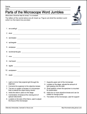
- Microscope Parts Worksheet Crossword Puzzle Answers
- Microscope Diagram Worksheet
- Microscope Parts Quiz Worksheet
- Microscope Mania Unit Review Crossword Puzzle
- Cell Microscope Lab Worksheet
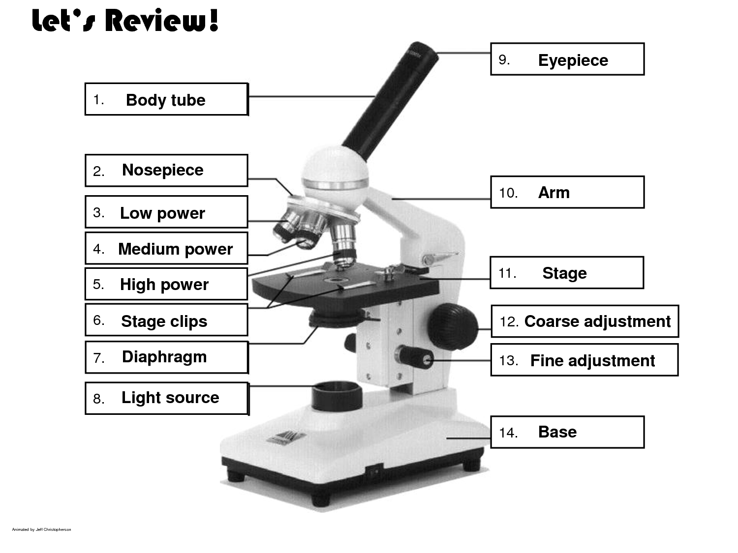
 Compound Light Microscope Parts Worksheet
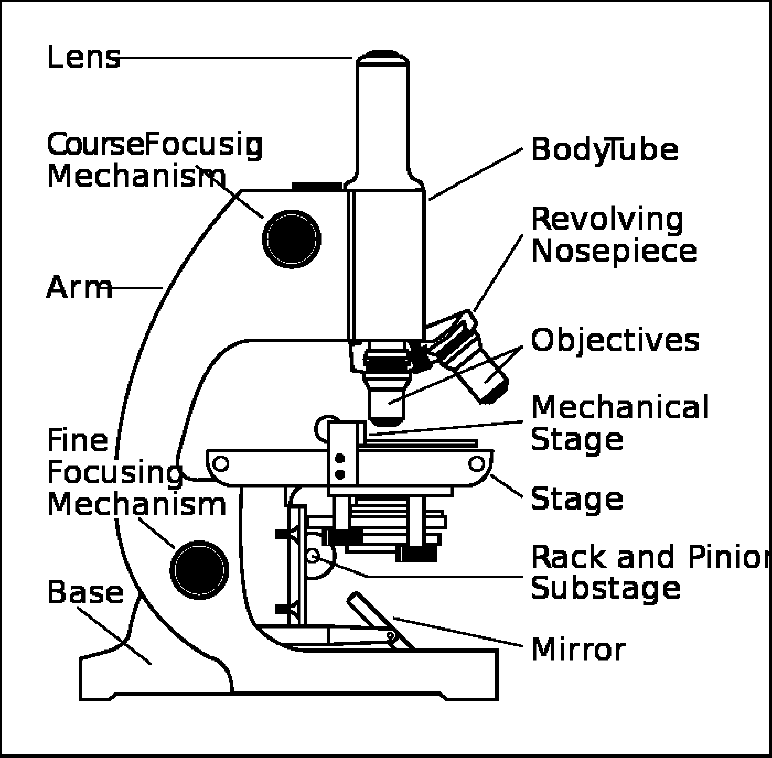
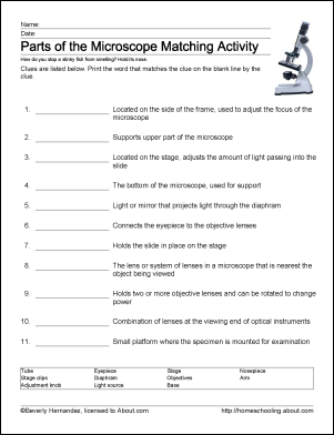
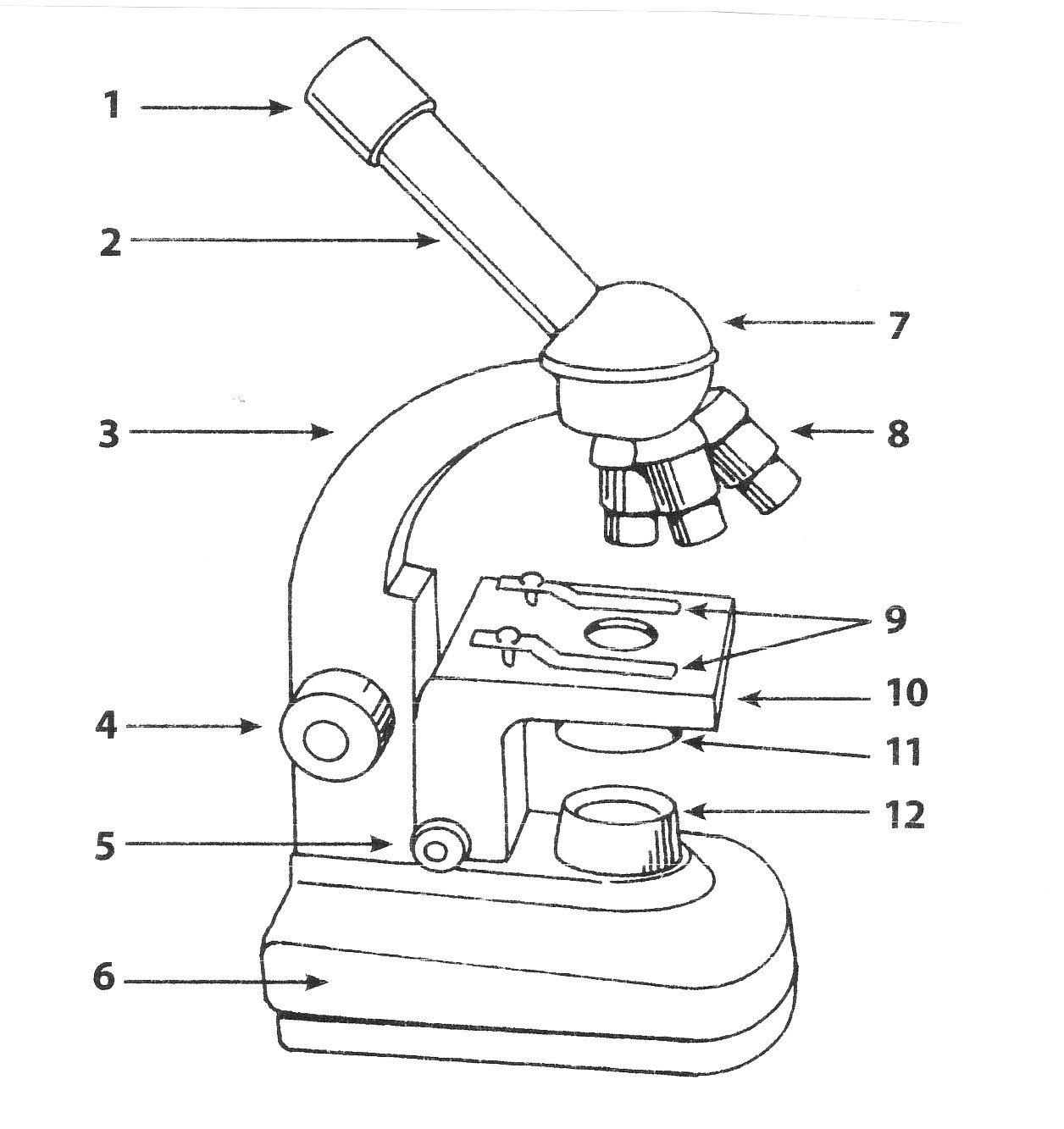
 Microscope Parts Worksheet
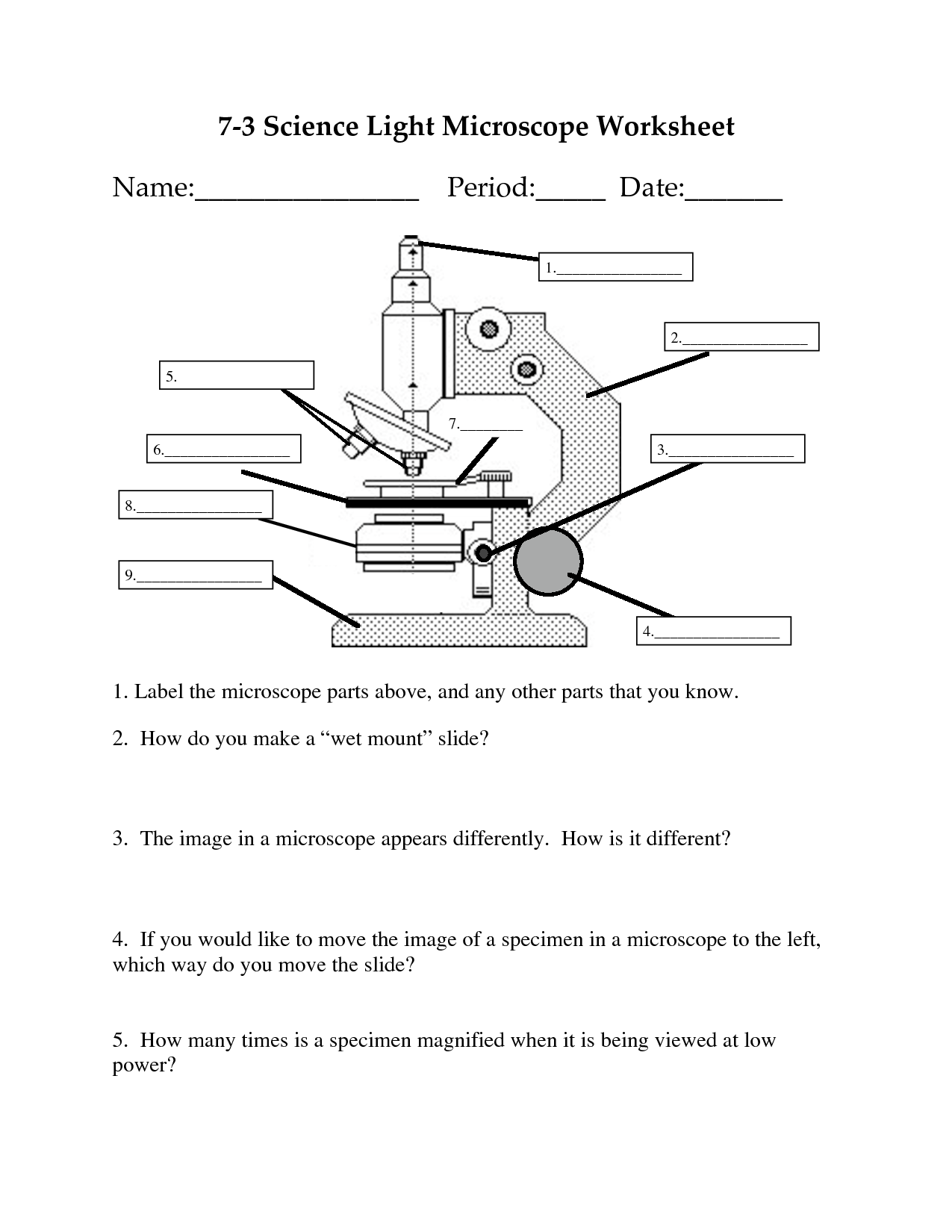
 Label Microscope Parts Worksheet
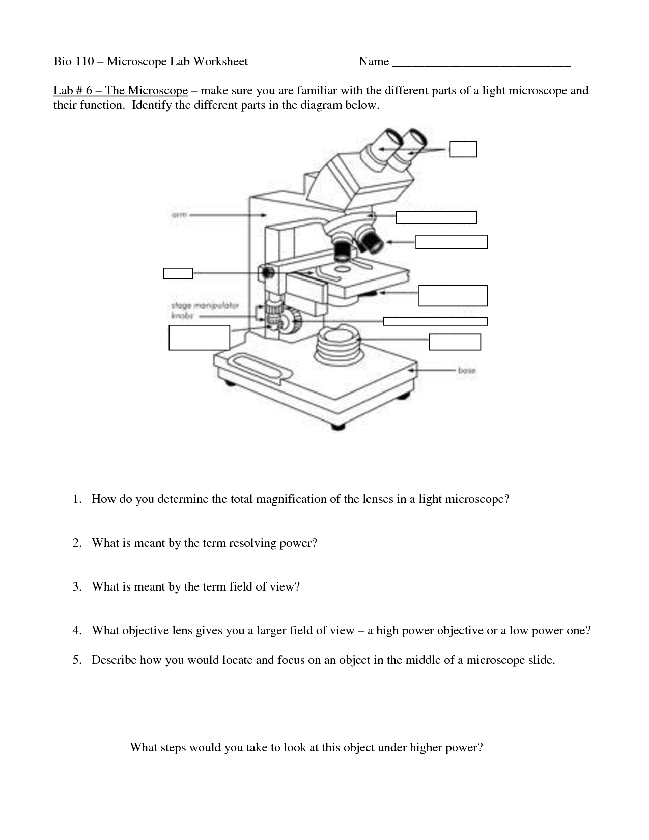
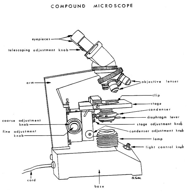
 Compound Microscope Worksheet
 Microscope Parts Worksheet Answers

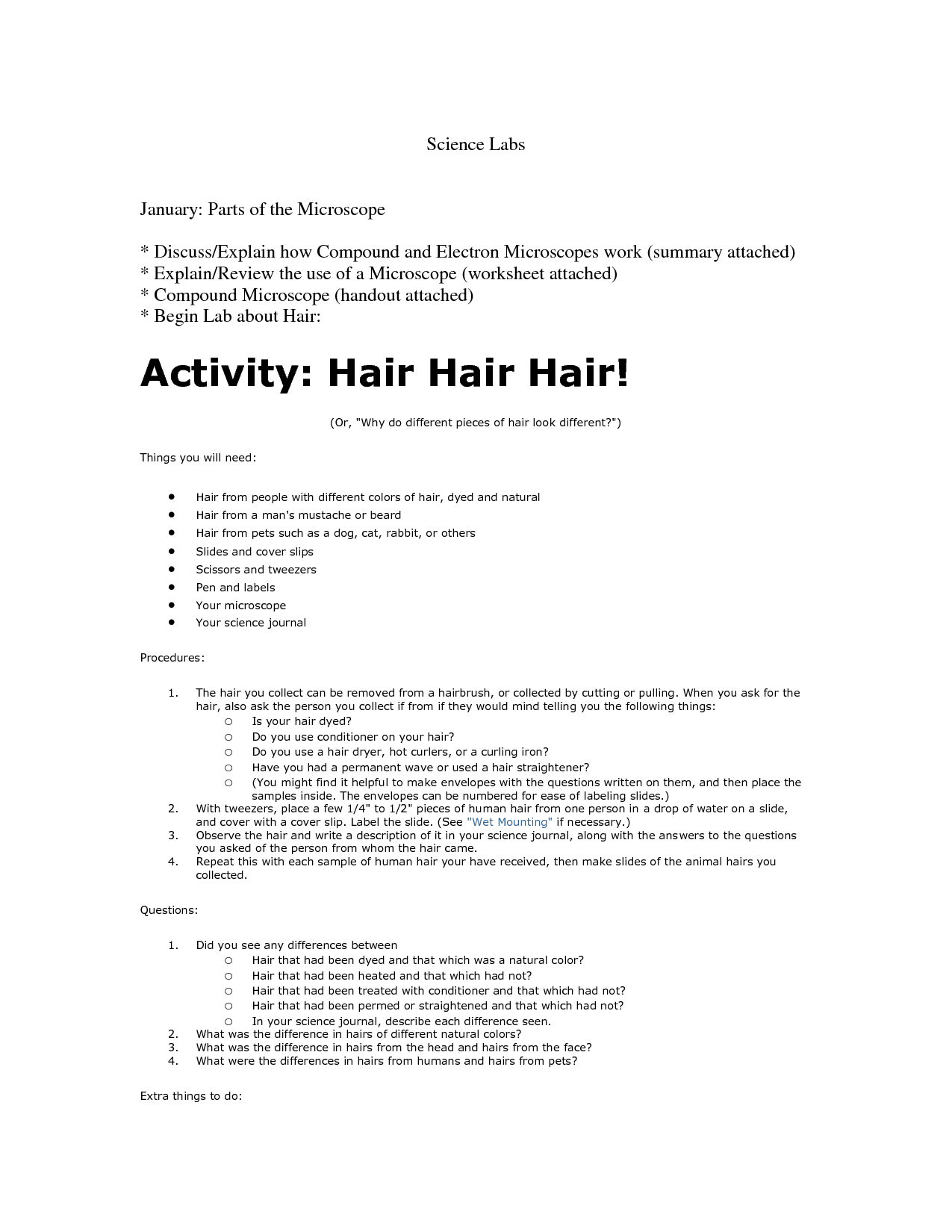
 Microscope Lab Worksheet Answers
 Microscope Parts Worksheet Answers
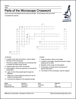
 Microscope Parts Worksheet Crossword Puzzle Answers
 Microscope Parts Worksheet
 Microscope Parts Worksheet Answers
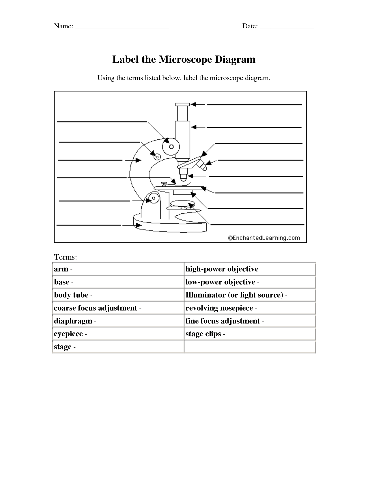
 Microscope Diagram Worksheet
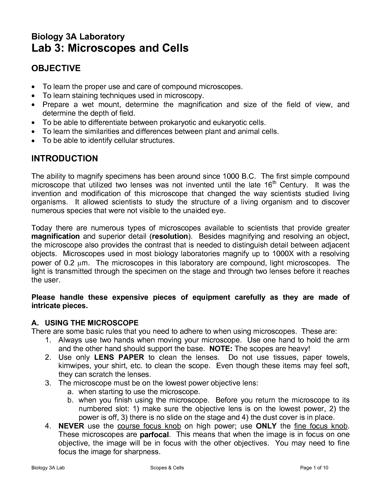
 Microscope Lab Worksheet Answers
 Microscope Parts Quiz Worksheet

 Microscope Mania Unit Review Crossword Puzzle
 Cell Microscope Lab Worksheet
More Other Worksheets
Kindergarten Worksheet My RoomSpanish Verb Worksheets
Cooking Vocabulary Worksheet
DNA Code Worksheet
Meiosis Worksheet Answer Key
Art Handouts and Worksheets
7 Elements of Art Worksheets
All Amendment Worksheet
Symmetry Art Worksheets
Daily Meal Planning Worksheet
What is a microscope?
A microscope is a scientific instrument that magnifies tiny objects or organisms that are too small to be seen by the naked eye, allowing for detailed examination and study of their structures.
What are the main parts of a microscope?
The main parts of a microscope typically include the eyepiece, objective lenses, stage, focus knobs, illuminator or light source, and the base. The eyepiece is where you look through to see the specimen. The objective lenses are used to magnify the specimen. The stage is where the specimen is placed for observation. The focus knobs are used to adjust the focus of the specimen. The illuminator or light source provides light to illuminate the specimen. Lastly, the base provides support and stability for the microscope.
How does a microscope work?
A microscope works by using lenses to magnify and focus light on a specimen, allowing you to see small details that are not visible to the naked eye. Light passes through the specimen and the lens system magnifies the image, which can be observed through the eyepiece or a camera. The power of the magnification depends on the type of lenses used in the microscope.
What is the difference between a compound microscope and a stereo microscope?
A compound microscope uses two sets of lenses (objective and eyepiece) to magnify small, thinly sliced specimens in 2D, providing high magnification and resolution for viewing cells and microorganisms. On the other hand, a stereo microscope, also known as a dissecting microscope, uses two separate optical pathways for each eyepiece to create a three-dimensional image of larger, solid specimens with lower magnification but greater depth perception. Stereo microscopes are ideal for tasks like dissection, soldering, and detailed inspection of larger objects in 3D.
What is magnification in microscopy?
Magnification in microscopy refers to the ability of a microscope to increase the apparent size of an object being viewed. It is achieved by multiplying the size of the object by a factor, allowing for the visualization of fine details that may not be visible to the naked eye. Magnification is typically represented as a ratio or a factor, such as 10x (10 times magnification), indicating how much larger the object appears compared to its actual size.
What is resolution in microscopy?
Resolution in microscopy refers to the ability of a microscope to distinguish between two closely spaced objects as separate entities. It is a measure of how sharp or clear the image produced by the microscope is, and is influenced by factors such as wavelength of light used, numerical aperture of the lens, and the quality of the optics. High resolution microscopes are capable of producing detailed and crisp images, while low resolution microscopes may not be able to distinguish fine details.
What is the working distance in microscopy?
Working distance in microscopy refers to the distance between the objective lens of a microscope and the specimen being viewed. It is important in determining the amount of space available for various manipulations such as sample preparation and making adjustments to the focus. A longer working distance allows for more flexibility in sample handling and is especially useful when studying thick samples or conducting experiments that require modifications to the specimen during observation.
What is the field of view in microscopy?
The field of view in microscopy refers to the area of the specimen that is visible through the microscope lens at a given magnification. It is the diameter of the circular image that you see when looking through the eyepiece of a microscope. A larger field of view allows you to see a larger portion of the specimen at once, whereas a smaller field of view provides higher magnification but only shows a smaller area of the specimen.
What is the importance of proper slide preparation in microscopy?
Proper slide preparation is crucial in microscopy as it ensures accurate and clear results. It helps to minimize distortions, artifacts, and contamination that could affect the quality of the image. By properly mounting and staining the specimen on the slide, it allows for better visualization of cells, tissues, or microorganisms under the microscope, aiding in accurate analysis and diagnosis in various fields such as medicine, biology, and material science. Ultimately, the quality of the slide preparation directly impacts the reliability and validity of the microscopic observations and findings.
What are some common applications of microscopy in research and everyday life?
Microscopy is used in research for various applications such as examining cell structures, studying microorganisms, analyzing material properties at the nanoscale, and observing chemical reactions at the molecular level. In everyday life, microscopy is used in fields like medicine for diagnosing diseases, in forensics for analyzing evidence, in environmental science for studying pollutants, in quality control for assessing product integrity, and in the food industry for inspecting food products for safety and quality.


Have something to share?
Who is Worksheeto?
At Worksheeto, we are committed to delivering an extensive and varied portfolio of superior quality worksheets, designed to address the educational demands of students, educators, and parents.


Comments