Lipid Coloring Worksheet
If you are searching for a practical and engaging way to teach your biology students about lipids, then look no further than the lipid coloring worksheet. This worksheet is designed to provide an interactive learning experience, allowing students to visually understand the structure and function of lipids, their classification, and their role in the human body. Whether you are a high school teacher or a college professor, this worksheet offers a valuable tool to help your students grasp the complexities of lipids.
Table of Images 👆
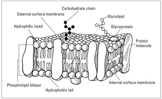
 Fluid Mosaic Model of the Cell Membrane

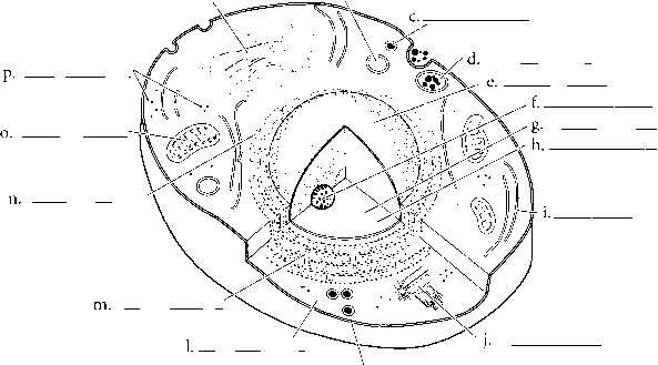
 Cell Membrane Diagram Labeled

 Elvis Presley Coloring Pages

 Rabbit Coloring Pages

 Christmas Santa Coloring Page
 Cell Organelles Coloring

 Doraemon Coloring Pages

 Fat Cat Coloring Pages
More Other Worksheets
Kindergarten Worksheet My RoomSpanish Verb Worksheets
Cooking Vocabulary Worksheet
DNA Code Worksheet
Meiosis Worksheet Answer Key
Art Handouts and Worksheets
7 Elements of Art Worksheets
All Amendment Worksheet
Symmetry Art Worksheets
Daily Meal Planning Worksheet
What is the purpose of lipid coloring in scientific research?
Lipid coloring in scientific research is done to visualize and track lipids within cells and tissues. By using specific dyes or fluorescent probes, researchers can study lipid metabolism, distribution, and interactions within biological systems. This technique helps to understand the roles of lipids in cellular processes, disease mechanisms, and drug development, providing valuable insights into various biological phenomena.
What are the different methods used to color lipids?
Some methods used to color lipids include staining with dyes like Sudan III or Oil Red O, which can impart a red color to lipids; using iodine to visualize lipids as they turn brown or black when mixed with iodine in a solution; and utilizing fluorescent dyes or probes that can specifically bind to lipids and emit fluorescence, allowing for their visualization under a microscope or other imaging techniques. These methods can help researchers and scientists identify and study the distribution and composition of lipids in various biological samples.
How does lipid coloring help in visualizing lipid distribution in cells or tissues?
Lipid coloring allows for the visualization of lipid distribution in cells or tissues by staining lipids with specific dyes or fluorescent probes that specifically bind to lipids. These dyes or probes help researchers to distinguish and track the location and quantity of lipids within cells or tissues under a microscope, providing valuable insights into lipid distribution, dynamics, and interactions within biological systems.
What are some commonly used lipid dyes or stains?
Some commonly used lipid dyes or stains include Oil Red O, Sudan Black B, Nile Red, and BODIPY dyes, which can be used to visualize and stain lipid droplets in cells and tissues for microscopy and imaging studies. These dyes have unique fluorescence properties that allow researchers to selectively label lipid-rich regions in biological samples.
How does lipid coloring distinguish between different lipid species?
Lipid coloring, such as with lipid dyes or stains, distinguishes between different lipid species based on their unique chemical properties and compositions. These dyes specifically bind to certain lipid components, such as fatty acids or polar head groups, producing distinct color patterns or intensities that help to visually differentiate between various lipid species under a microscope or in other analytical techniques like chromatography. By exploiting the differences in lipid compositions, lipid coloring can provide valuable information about the distribution and abundance of specific lipid species within cells or tissues.
How is the intensity of lipid coloring related to lipid concentration?
The intensity of lipid coloring is directly related to the lipid concentration, meaning that as the lipid concentration increases, the intensity of the coloring also increases. This relationship is due to the higher amount of lipids present in the sample leading to a more pronounced color reaction with the specific lipid staining or dye being used, resulting in a darker or more vibrant color. Essentially, the more lipids present, the stronger the coloring will be, allowing for easier visual detection and quantification of lipids in a sample.
Can lipid coloring be used to detect lipid abnormalities or imbalances?
No, lipid coloring cannot be used to detect lipid abnormalities or imbalances. Lipid coloring is used to visualize lipids in tissues or cells for research purposes, but it does not provide information on lipid levels or abnormalities. To detect lipid abnormalities or imbalances, specific lipid tests such as cholesterol and triglyceride measurements are typically performed.
Are there any limitations or challenges associated with lipid coloring techniques?
Yes, there are limitations and challenges associated with lipid coloring techniques. One limitation is the lack of specificity in some staining methods, which may result in non-specific binding and background interference. Additionally, certain lipid classes may not be effectively visualized using traditional staining approaches, leading to incomplete or inaccurate characterization. Challenges also include the variability in lipid composition between samples, the need for specialized equipment and expertise, and the potential for sample degradation during staining procedures. Overall, careful optimization and validation of lipid coloring techniques are essential to overcome these limitations and challenges in lipid analysis.
Can lipid coloring be used in live cell imaging?
Yes, lipid coloring can be used in live cell imaging to visualize the distribution and dynamics of lipids within cells. Fluorescent lipid dyes or probes can selectively label different lipid species, allowing researchers to track their movement, localization, and interactions with other cellular components in real-time. This technique is valuable for studying various lipid-related processes, including membrane trafficking, lipid metabolism, and signaling pathways, in living cells.
Are there any emerging technologies or advancements in lipid coloring techniques?
Yes, there are emerging technologies and advancements in lipid coloring techniques. For instance, advanced microscopy techniques like fluorescence microscopy coupled with lipid-specific dyes or probes are being developed to visualize lipids in cells and tissues with high spatial resolution. Additionally, researchers are exploring new types of lipid dyes and probes that can selectively target specific lipid species or organelles within cells, providing more detailed insights into lipid dynamics and functions. These advancements in lipid coloring techniques are helping to further our understanding of the roles that lipids play in various cellular processes and diseases.


Have something to share?
Who is Worksheeto?
At Worksheeto, we are committed to delivering an extensive and varied portfolio of superior quality worksheets, designed to address the educational demands of students, educators, and parents.


Comments