Human Heart Diagram Worksheet
The human heart diagram worksheet is a valuable educational resource that offers a comprehensive visual representation of the intricacies of the human heart. Designed for students, teachers, and medical professionals alike, this worksheet provides an in-depth exploration of the various components and functions of the heart. With clear and detailed illustrations, learners can easily grasp the anatomy and physiology of this vital organ.
Table of Images 👆
- Label Heart Diagram Worksheet
- Human Body Systems Labeling Worksheet
- Circulatory System Worksheets
- Heart Anatomy Diagram Worksheet
- Human Heart Diagram Coloring
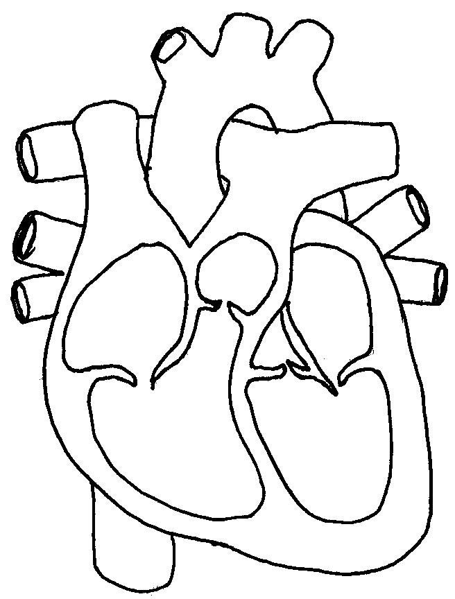
- Blank Heart Diagram Blood Flow
- Blank Human Body Outline Template
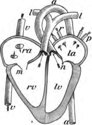
- Labeled Heart Diagram
- Circulatory System Heart Diagram Worksheet
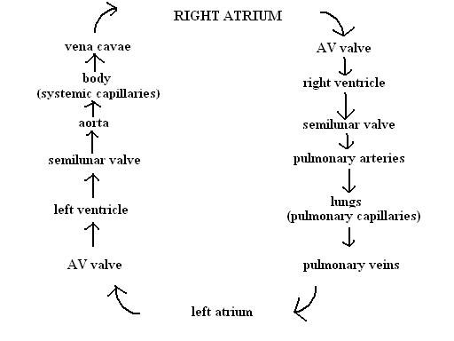
- Circulatory System Flow Chart
- Gas Exchange Respiratory System Diagram
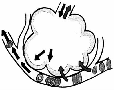
- In the Right Atrium of Heart Blood Flow through Starting
- Circulatory System Heart Diagram
- Electric Shock Effects On Human Body
- Cartoon Facial Expressions
- Connective Tissue Types and Functions

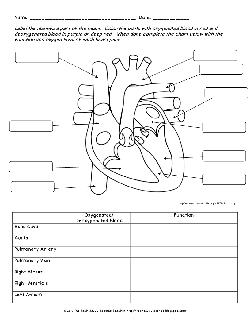
 Label Heart Diagram Worksheet
 Human Body Systems Labeling Worksheet
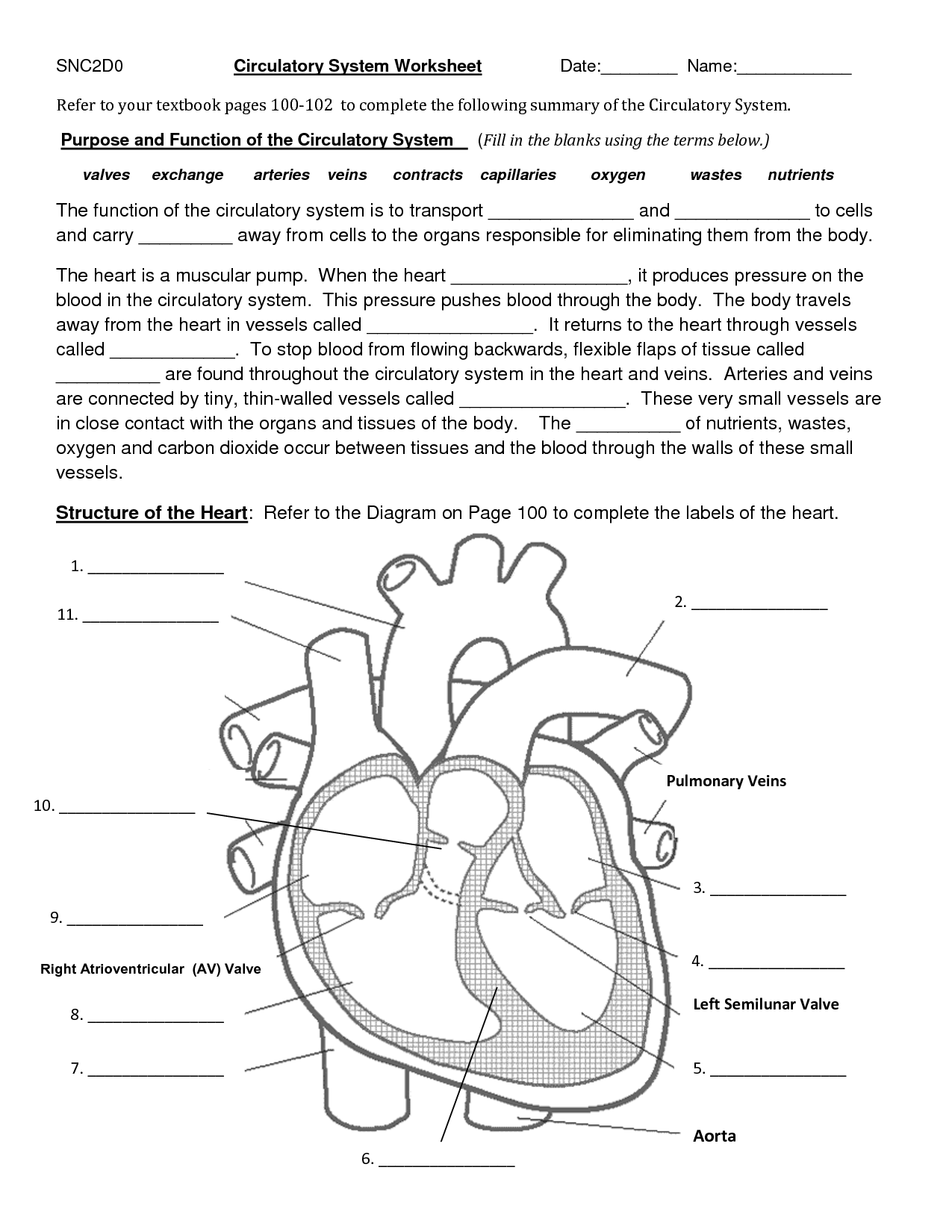
 Circulatory System Worksheets
 Heart Anatomy Diagram Worksheet
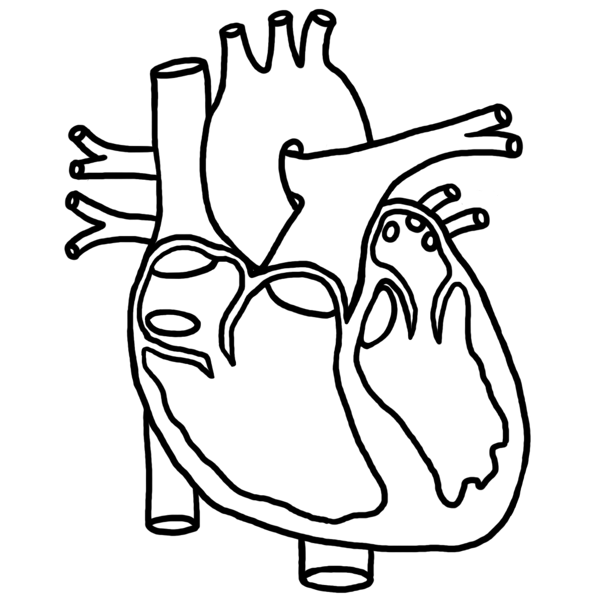
 Human Heart Diagram Coloring
 Blank Heart Diagram Blood Flow
 Human Heart Diagram Coloring

 Blank Human Body Outline Template

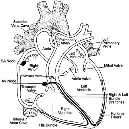
 Labeled Heart Diagram
 Circulatory System Heart Diagram Worksheet
 Circulatory System Flow Chart
 Gas Exchange Respiratory System Diagram
 In the Right Atrium of Heart Blood Flow through Starting
 Circulatory System Heart Diagram
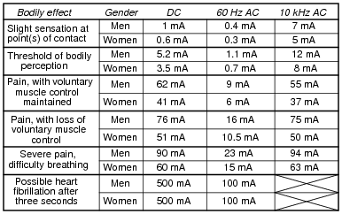
 Electric Shock Effects On Human Body
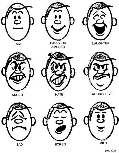
 Cartoon Facial Expressions

 Connective Tissue Types and Functions
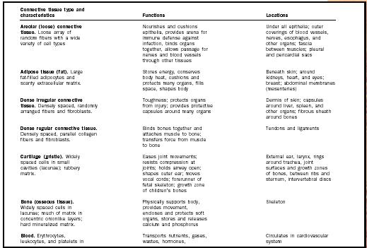
 Connective Tissue Types and Functions
More Other Worksheets
Kindergarten Worksheet My RoomSpanish Verb Worksheets
Cooking Vocabulary Worksheet
DNA Code Worksheet
Meiosis Worksheet Answer Key
Art Handouts and Worksheets
7 Elements of Art Worksheets
All Amendment Worksheet
Symmetry Art Worksheets
Daily Meal Planning Worksheet
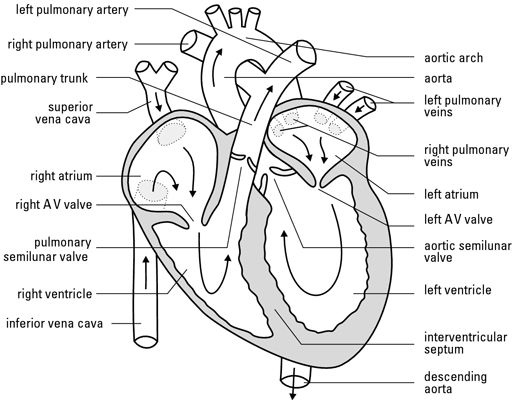
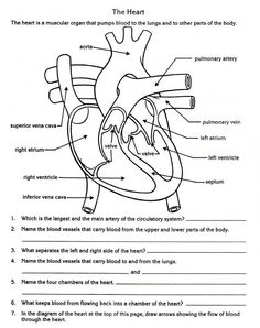
Label the four chambers of the human heart.
The four chambers of the human heart are the left atrium, right atrium, left ventricle, and right ventricle.
Identify the main blood vessels connected to the heart.
The main blood vessels connected to the heart are the aorta, which carries oxygenated blood from the heart to the rest of the body, and the pulmonary arteries, which carry deoxygenated blood from the heart to the lungs for oxygenation. Additionally, the superior and inferior vena cava are the main veins that return deoxygenated blood from the body back to the heart.
Describe the function of the valves in the heart.
Valves in the heart function to ensure proper blood flow by allowing blood to flow in one direction while preventing backflow. The two main types of valves in the heart, atrioventricular valves, and semilunar valves, open and close in coordination with the heart's contractions, regulating the flow of blood from the atria to the ventricles and out of the heart to the pulmonary artery and aorta, respectively. This mechanism helps maintain the efficiency and effectiveness of the heart's pumping action, ensuring adequate circulation throughout the body.
Explain the purpose of the coronary arteries.
The coronary arteries are responsible for supplying the heart muscle with oxygen and nutrients that are essential for proper functioning. They play a crucial role in ensuring that the heart receives an adequate blood supply, allowing it to pump efficiently and without interruption. If the coronary arteries become blocked or constricted, it can lead to serious conditions such as coronary artery disease, heart attacks, or other cardiovascular complications.
Identify the location and function of the sinoatrial (SA) node.
The sinoatrial (SA) node is located in the right atrium of the heart. It serves as the natural pacemaker of the heart, generating electrical impulses that initiate each heartbeat and coordinate the heart's rhythm by regulating the heart rate.
Label the major blood vessels that deliver oxygenated and deoxygenated blood to and from the heart.
The major blood vessels that deliver oxygenated blood to the heart are the pulmonary veins, which carry oxygenated blood from the lungs to the left atrium, and the major blood vessel that delivers deoxygenated blood to the heart is the superior and inferior vena cava, which bring deoxygenated blood from the body back to the right atrium.
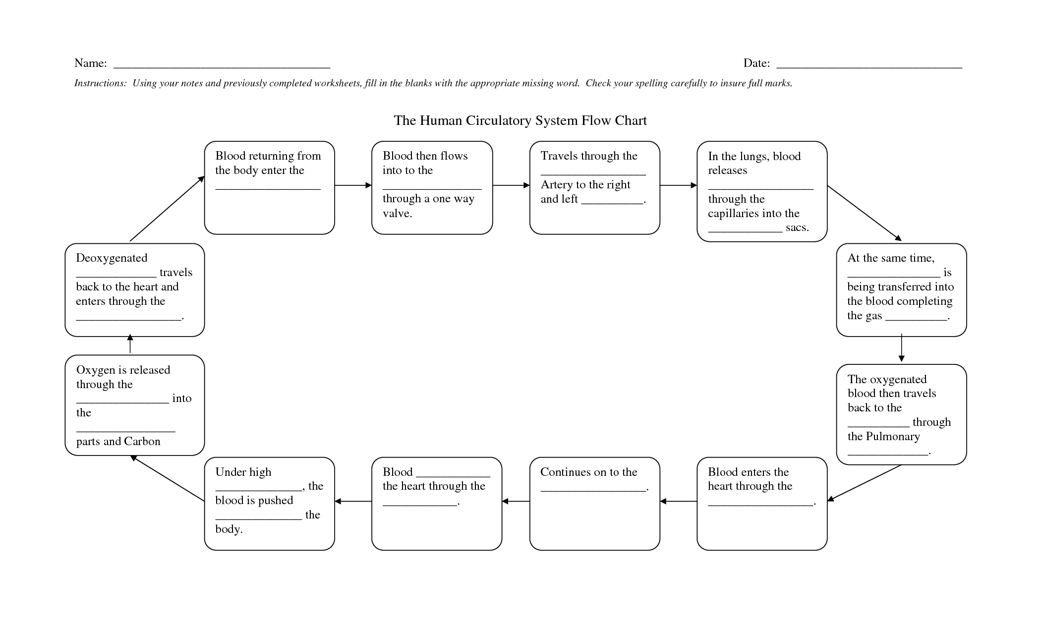
Describe the pathway of blood flow through the heart.
Blood enters the heart through the right atrium from the body, then flows into the right ventricle, where it is pumped to the lungs for oxygenation. After picking up oxygen from the lungs, the blood returns to the heart through the left atrium, then enters the left ventricle, which pumps the oxygen-rich blood out to the rest of the body. This continuous cycle of blood flow through the heart is essential for distributing oxygen and nutrients to the body's tissues and organs.
Identify the layers of the heart wall.
The layers of the heart wall are the outermost epicardium, the middle myocardium responsible for the heart's pumping action, and the inner endocardium which lines the chambers of the heart.
Explain the role of the cardiac muscle in the heart.
The cardiac muscle in the heart plays a crucial role in pumping and circulating blood throughout the body. It contracts and relaxes in a coordinated manner to ensure that blood is efficiently pumped out of the heart chambers, maintaining blood flow to the organs and tissues. The continuous and rhythmic contraction of the cardiac muscle generates the necessary force to propel the blood through the blood vessels, providing oxygen and nutrients to the cells while removing waste products. This coordinated action is essential for the heart to effectively fulfill its function as the body's main organ for circulation.
Describe the function of the heart as a vital organ in the circulatory system.
The heart serves as the central pump of the circulatory system, responsible for pumping oxygen-rich blood to all tissues and organs of the body and collecting oxygen-poor blood to be sent to the lungs for oxygenation. It maintains blood flow through the network of blood vessels, ensuring nutrients, hormones, and waste products are transported to and from cells. The heart's rhythmic contractions, controlled by electrical impulses, enable the circulation of blood, providing essential oxygen and nutrients to sustain life and remove metabolic waste products.


Have something to share?
Who is Worksheeto?
At Worksheeto, we are committed to delivering an extensive and varied portfolio of superior quality worksheets, designed to address the educational demands of students, educators, and parents.


Comments