Eye Anatomy Worksheet
Are you a student or an educator looking for an engaging and informative way to learn about the intricate details of the human eye? Look no further! Our eye anatomy worksheet provides a comprehensive overview of the various structures and functions of the eye, making it the perfect learning tool for biology or anatomy enthusiasts.
Table of Images 👆
- Human Eye Diagram Unlabeled
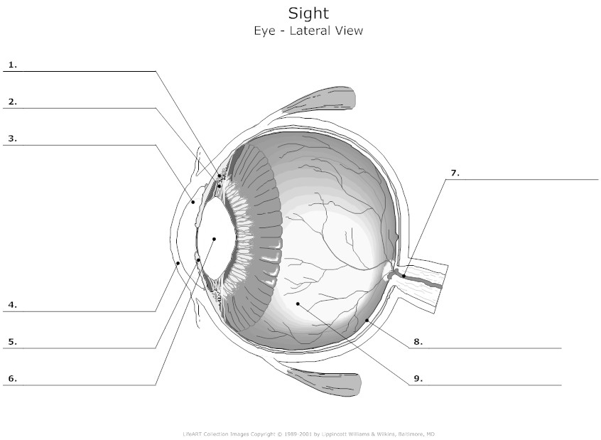
- Human Eye Anatomy Diagram Worksheet
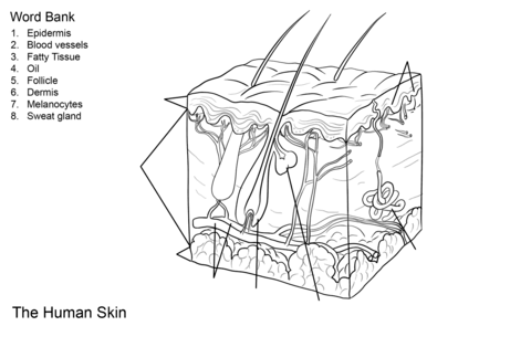
- Human Skin Anatomy Worksheet Coloring Page
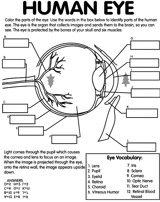
- Human Eye Diagram
- Anatomy and Physiology Eye Diagram Label
- Human Eye Worksheet Answers
- Human Skeleton Bones Worksheet
- Human Eye Coloring Page
- Nervous System Anatomy and Physiology Worksheets
- Frog Dissection Worksheet Diagram
- Human Ear Diagram Unlabeled
- Human Brain Diagram Outline
- Human Heart Diagram Unlabeled
 Human Eye Diagram Unlabeled
 Human Eye Anatomy Diagram Worksheet
 Human Skin Anatomy Worksheet Coloring Page
 Human Eye Diagram

 Anatomy and Physiology Eye Diagram Label

 Human Eye Worksheet Answers
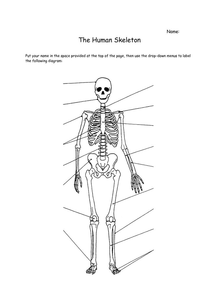
 Human Skeleton Bones Worksheet
 Human Eye Coloring Page
 Nervous System Anatomy and Physiology Worksheets

 Frog Dissection Worksheet Diagram
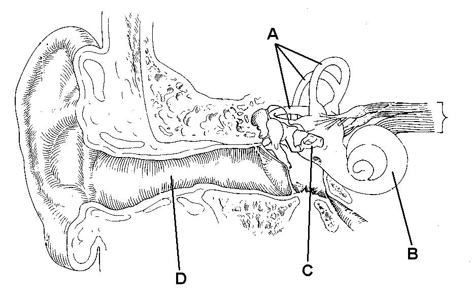
 Human Ear Diagram Unlabeled
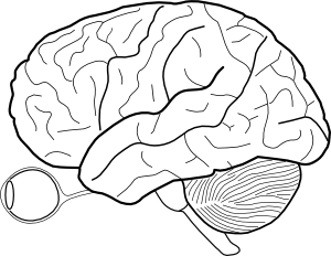
 Human Brain Diagram Outline
 Human Heart Diagram Unlabeled

 Human Heart Diagram Unlabeled

 Human Heart Diagram Unlabeled
More Other Worksheets
Kindergarten Worksheet My RoomSpanish Verb Worksheets
Cooking Vocabulary Worksheet
DNA Code Worksheet
Meiosis Worksheet Answer Key
Art Handouts and Worksheets
7 Elements of Art Worksheets
All Amendment Worksheet
Symmetry Art Worksheets
Daily Meal Planning Worksheet
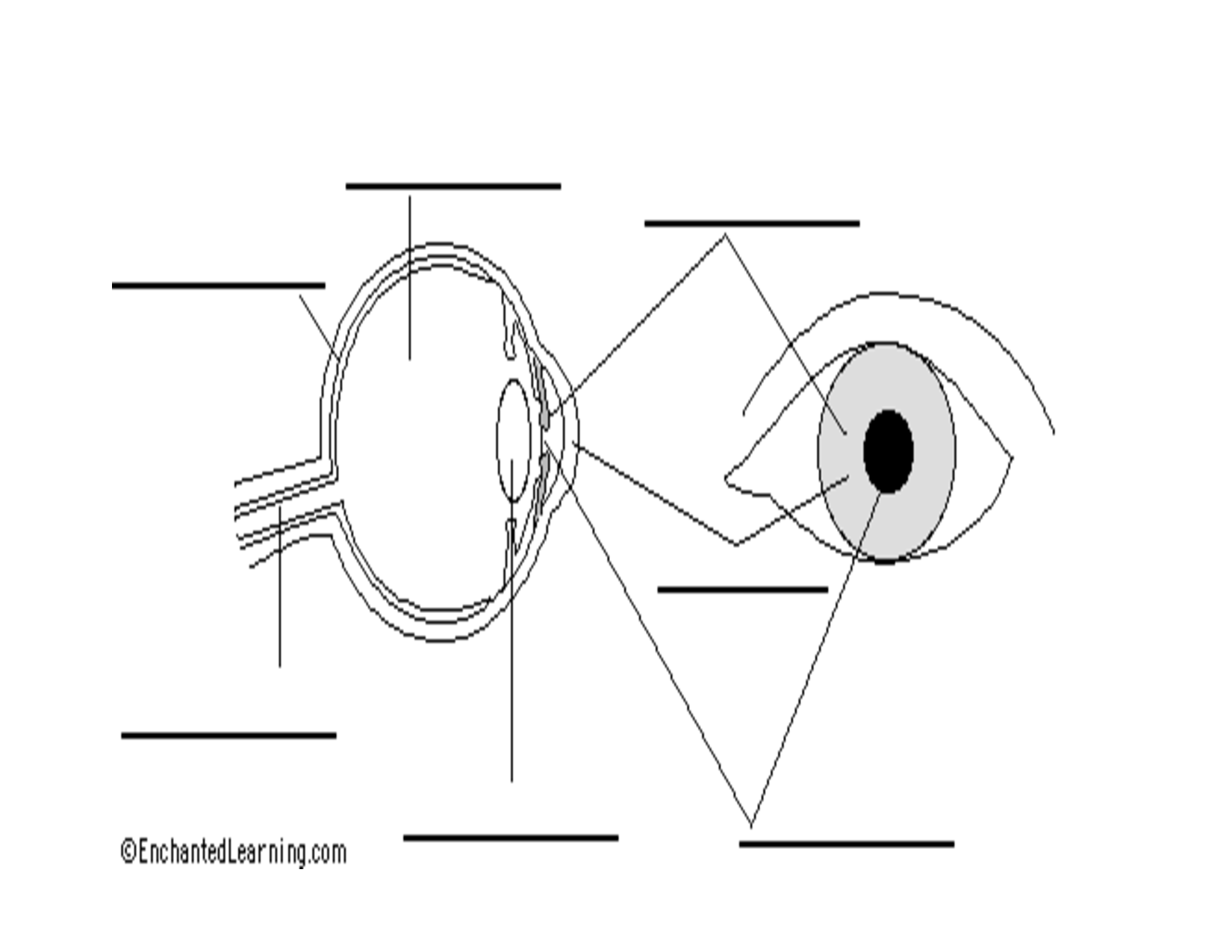
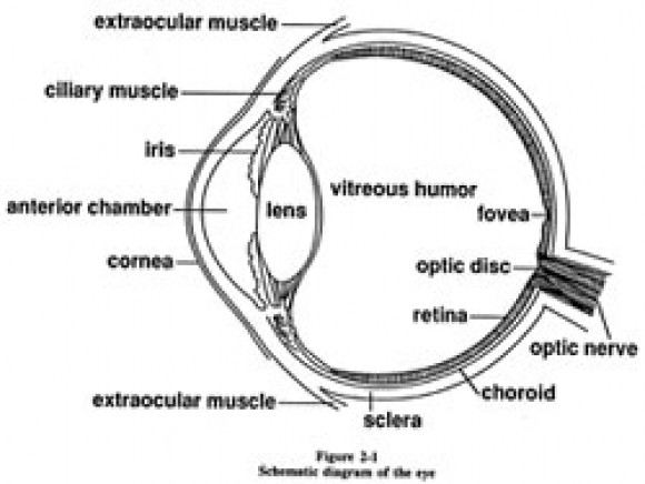
What is the purpose of the cornea?
The purpose of the cornea is to act as a transparent, protective outer covering of the eye that helps to focus light entering the eye onto the retina, enabling clear vision.
What are the three main layers of the eyeball?
The three main layers of the eyeball are the outer layer called the sclera and cornea, the middle layer known as the choroid, ciliary body, and iris, and the inner layer comprising the retina, which contains the light-sensitive cells that capture and process visual information.
What is the function of the lens?
The lens in the eye helps to focus incoming light onto the retina at the back of the eye, which then converts the light into electrical signals that are sent to the brain for visual interpretation.
Where is the iris located and what is its role?
The iris is located in the eye between the cornea and the lens. Its role is to regulate the amount of light entering the eye by adjusting the size of the pupil. The iris contains muscles that contract or relax in response to light intensity, making the pupil larger in dim light and smaller in bright light, thus controlling how much light reaches the retina at the back of the eye.
What are the three types of cones found in the retina and what do they do?
The three types of cones found in the retina are red cones, green cones, and blue cones. These cones are responsible for color vision and help us perceive different wavelengths of light. Red cones are most sensitive to long wavelengths of light, green cones to medium wavelengths, and blue cones to short wavelengths, allowing us to see a wide range of colors in our environment.
What is the purpose of the optic nerve?
The optic nerve carries visual information from the retina in the eye to the brain, allowing us to perceive and process visual stimuli such as images, colors, and shapes. It serves as the primary pathway for transmitting visual signals and plays a crucial role in our ability to see and interpret the world around us.
What is the function of the ciliary body?
The ciliary body is responsible for producing aqueous humor, a clear fluid that nourishes and maintains the shape of the eye. It also plays a crucial role in controlling the shape of the lens and helping the eye focus on objects at different distances, known as accommodation.
Where are the rods located and what is their role?
Rods are located in the retina of the eye, specifically in the outer layer known as the peripheral retina. Their role is to detect low levels of light, enabling night vision and providing the ability to see in dimly lit environments. Rods are particularly sensitive to light, but they do not distinguish color.
What is the purpose of the vitreous humor?
The purpose of the vitreous humor is to maintain the shape of the eye, provide a clear pathway for light entering the eye, and support the structures within the eye such as the lens and retina. It also helps maintain intraocular pressure and contributes to the overall structural integrity of the eye.
What is the function of the fovea centralis?
The function of the fovea centralis is to provide the sharpest vision in the eye by containing a high density of cone cells, which are responsible for detailed color vision in well-lit conditions. This area of the retina is essential for tasks that require precise visual acuity, such as reading, recognizing faces, and focusing on fine details.


Have something to share?
Who is Worksheeto?
At Worksheeto, we are committed to delivering an extensive and varied portfolio of superior quality worksheets, designed to address the educational demands of students, educators, and parents.


Comments