Anatomy Muscle Labeling Worksheet
The anatomy muscle labeling worksheet is an educational tool designed to help students better understand the structure and function of different muscles in the human body. By providing a visual representation of muscle groups and their corresponding labels, this worksheet allows learners to engage in a hands-on approach to studying anatomy and physiology.
Table of Images 👆
- Blank Head and Neck Muscles Diagram
- Skeletal System Diagram Worksheet
- Muscular System Muscle Anatomy
- Bone Anatomy Labeling Worksheets
- Medial Condyle of Femur Bone
- Nervous System Spinal Cord Diagram
- Body Tissues Worksheet Answers
- Sensory and Motor Neurons Spinal Cord Anatomy
- Small Intestine Wall Diagram
- Blank Skeletal System Diagram
- Dogfish Shark External Anatomy Diagram
- Tibia and Fibula Diagram Unlabeled
- Muscular System Printable Worksheets
- Label Heart Diagram Worksheet
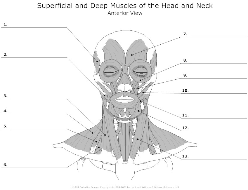
 Blank Head and Neck Muscles Diagram
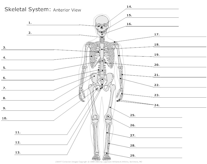
 Skeletal System Diagram Worksheet
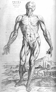
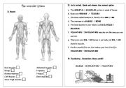
 Muscular System Muscle Anatomy
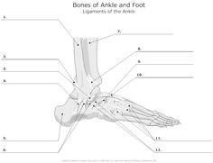
 Bone Anatomy Labeling Worksheets
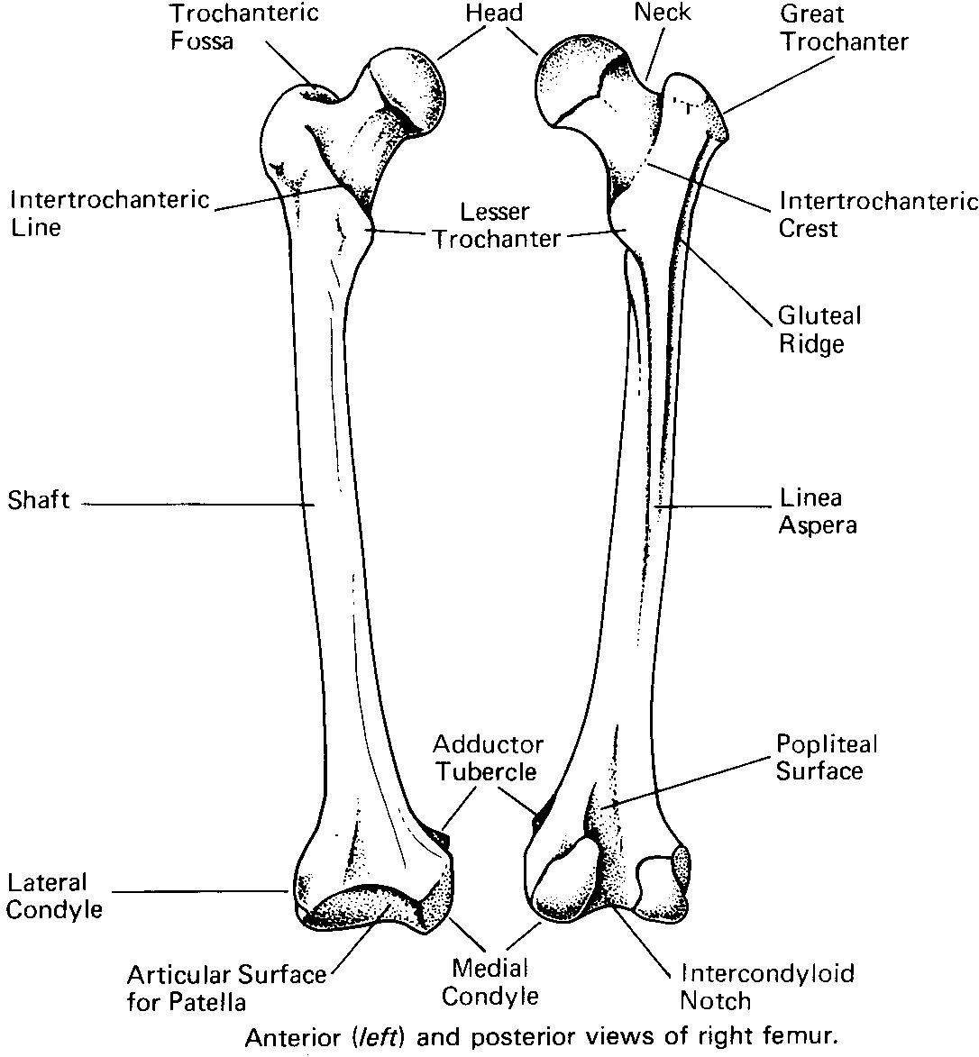
 Medial Condyle of Femur Bone
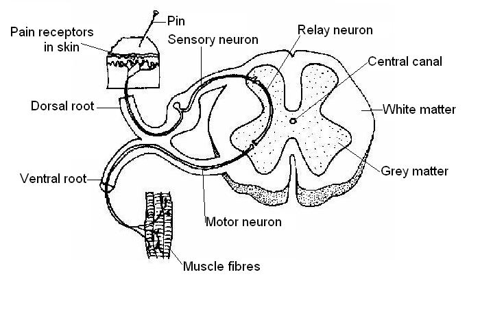
 Nervous System Spinal Cord Diagram
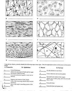
 Body Tissues Worksheet Answers
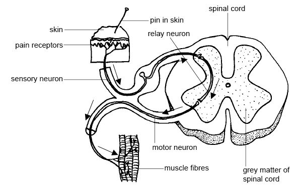
 Sensory and Motor Neurons Spinal Cord Anatomy
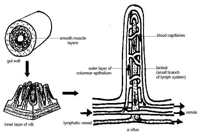
 Small Intestine Wall Diagram

 Blank Skeletal System Diagram
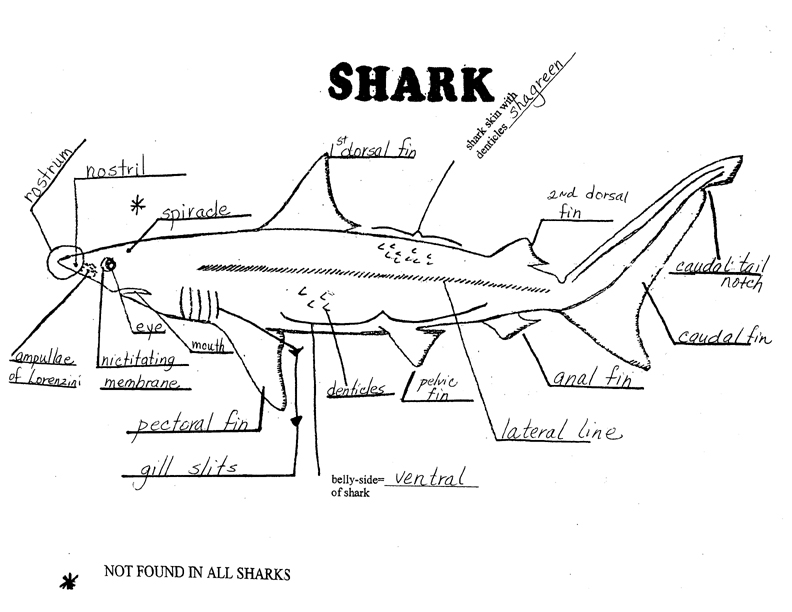
 Dogfish Shark External Anatomy Diagram
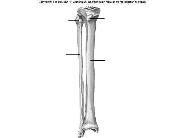
 Tibia and Fibula Diagram Unlabeled
 Muscular System Printable Worksheets

 Label Heart Diagram Worksheet
More Other Worksheets
Kindergarten Worksheet My RoomSpanish Verb Worksheets
Cooking Vocabulary Worksheet
DNA Code Worksheet
Meiosis Worksheet Answer Key
Art Handouts and Worksheets
7 Elements of Art Worksheets
All Amendment Worksheet
Symmetry Art Worksheets
Daily Meal Planning Worksheet
What is the origin of the biceps brachii muscle?
The biceps brachii muscle originates from two different points on the scapula (shoulder blade) - the long head originates from the supraglenoid tubercle above the shoulder joint, and the short head originates from the coracoid process, which is a protrusion on the front of the scapula.
What is the insertion of the trapezius muscle?
The insertion of the trapezius muscle is along the spine of the scapula and the acromion and spine of the clavicle. It plays a key role in scapular stabilization and movement, primarily shoulder elevation and retraction.
What is the action of the gastrocnemius muscle?
The action of the gastrocnemius muscle is to plantarflex the foot, meaning it helps to point the foot downwards or flex the ankle joint.
What is the innervation of the deltoid muscle?
The deltoid muscle is innervated by the axillary nerve, which arises from the posterior cord of the brachial plexus.
What is the function of the rectus abdominis muscle?
The rectus abdominis muscle functions to flex the trunk, tilt the pelvis, and stabilize the core. It is responsible for movements like bending forward, sitting up, and maintaining good posture. Additionally, the rectus abdominis muscle plays a key role in providing support and protection to the abdominal organs.
What is the name of the muscle responsible for flexing the hip joint?
The muscle responsible for flexing the hip joint is called the iliopsoas, which is a combination of the psoas major and the iliacus muscles. Together, they work to bring the thigh towards the abdomen in a movement known as hip flexion.
What is the origin of the triceps brachii muscle?
The triceps brachii muscle originates from three separate heads: the long head originates from the infraglenoid tubercle of the scapula, the lateral head originates from the posterior humerus above the radial groove, and the medial head originates from the posterior humerus below the radial groove.
What is the insertion of the pectoralis major muscle?
The pectoralis major muscle inserts into the intertubercular groove of the humerus, also known as the bicipital groove or the groove of the lesser tubercle on the anterior aspect of the humerus.
What is the action of the quadriceps femoris muscle?
The quadriceps femoris muscle is responsible for extending the knee joint and straightening the leg. It is the primary muscle involved in activities such as walking, running, jumping, and squatting.
What is the innervation of the gluteus maximus muscle?
The gluteus maximus muscle is primarily innervated by the inferior gluteal nerve, which is a branch of the sacral plexus.


Have something to share?
Who is Worksheeto?
At Worksheeto, we are committed to delivering an extensive and varied portfolio of superior quality worksheets, designed to address the educational demands of students, educators, and parents.


Comments