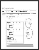
Abdominal Ultrasound Worksheet
An abdominal ultrasound worksheet is a helpful tool for healthcare professionals who perform ultrasounds on patients' abdominal regions. This worksheet provides a structured format for documenting important information and observations during the ultrasound examination. With its focus on the entity being examined and the subject of the ultrasound, this worksheet is designed to ensure accurate and comprehensive reporting, which is crucial for effective diagnosis and treatment.
Table of Images 👆
 Ultrasound Technologist Worksheets

 Abdominal Aorta Ultrasound Worksheet
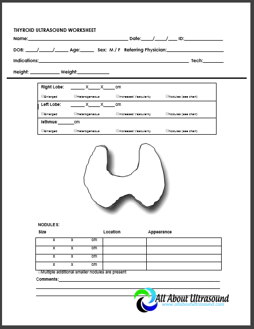
 Thyroid Ultrasound Worksheet
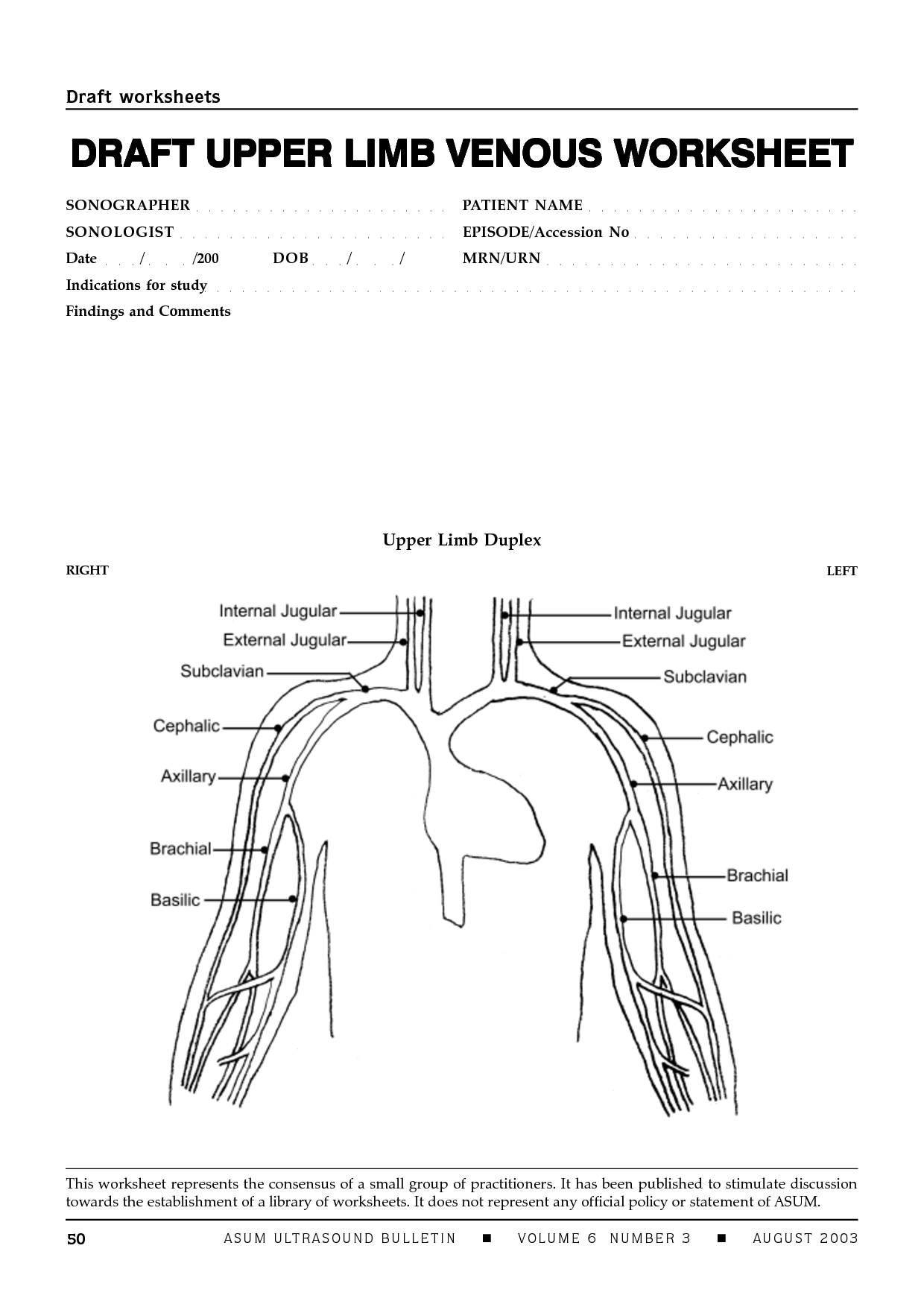
 Upper Extremity Arterial Ultrasound Worksheet

 Abdominal Aortic Dissection Ultrasound

 Graft Vascular Ultrasound Worksheet
 Graft Vascular Ultrasound Worksheet

 Graft Vascular Ultrasound Worksheet

 Graft Vascular Ultrasound Worksheet

 Graft Vascular Ultrasound Worksheet

 Graft Vascular Ultrasound Worksheet

 Graft Vascular Ultrasound Worksheet

 Graft Vascular Ultrasound Worksheet

 Graft Vascular Ultrasound Worksheet

 Graft Vascular Ultrasound Worksheet

 Graft Vascular Ultrasound Worksheet

 Graft Vascular Ultrasound Worksheet

 Graft Vascular Ultrasound Worksheet

 Graft Vascular Ultrasound Worksheet
More Other Worksheets
Kindergarten Worksheet My RoomSpanish Verb Worksheets
Cooking Vocabulary Worksheet
DNA Code Worksheet
Meiosis Worksheet Answer Key
Art Handouts and Worksheets
7 Elements of Art Worksheets
All Amendment Worksheet
Symmetry Art Worksheets
Daily Meal Planning Worksheet
What is the purpose of an abdominal ultrasound?
An abdominal ultrasound is used to visualize and assess the organs and structures within the abdomen, such as the liver, gallbladder, pancreas, kidneys, spleen, and abdominal aorta. It can help diagnose and monitor conditions such as gallstones, kidney stones, liver disease, abdominal masses, and abnormal fluid accumulation. Additionally, it can provide information about blood flow to these organs and detect potential abnormalities.
What are some common indications for an abdominal ultrasound?
Common indications for an abdominal ultrasound include evaluating organs such as the liver, gallbladder, pancreas, kidneys, and spleen for conditions like gallstones, liver disease, pancreatitis, kidney stones, and abdominal mass or tumors. It can also be used to investigate causes of abdominal pain, swelling, abnormal growths, or to monitor the progression of certain conditions like liver cirrhosis or kidney disease.
How is the patient prepared for an abdominal ultrasound?
The patient is prepared for an abdominal ultrasound by being asked to fast for several hours before the exam to ensure clear images of the organs in the abdominal area. They may also be asked to drink water before the test to have a full bladder, aiding in visualizing certain organs. Patients are typically instructed to wear loose, comfortable clothing and may need to remove any jewelry or metal objects that could interfere with the ultrasound.
What are the key components of the ultrasound machine used for abdominal imaging?
The key components of an ultrasound machine used for abdominal imaging include a transducer probe, which emits and receives ultrasound waves to create images, a monitor to display the images in real-time, a control panel to adjust settings and optimize image quality, a gel to enhance sound wave transmission, a computer system for processing and storing images, and a power source to operate the machine. These components work together to visualize abdominal organs and structures during imaging procedures.
What are the standard scanning techniques employed during an abdominal ultrasound?
The standard scanning techniques employed during an abdominal ultrasound include a combination of various approaches such as a subcostal scan, intercostal scan, and suprapubic scan. These techniques involve the use of high-frequency sound waves to create detailed images of the abdominal organs and structures by moving the ultrasound transducer across the skin surface while applying gel to ensure proper contact and visualization of the area of interest. The specific technique used will depend on the clinical indication, the anatomical region being assessed, and the patient's body habitus.
What are the typical measurements obtained during an abdominal ultrasound?
During an abdominal ultrasound, typical measurements obtained include the size of organs such as the liver, gallbladder, pancreas, kidneys, spleen, and bladder. Other measurements may include the thickness of the walls of organs like the gallbladder or bladder, the presence of any fluid collection or masses, and the diameter of blood vessels in the abdomen. These measurements help in assessing the structure, function, and overall health of the abdominal organs.
How is the liver evaluated during an abdominal ultrasound?
During an abdominal ultrasound, the liver is evaluated by examining its size, shape, and structure. The ultrasound technician will look for any abnormalities such as masses, cysts, or signs of liver disease. Blood flow within the liver can also be assessed using Doppler ultrasound to check for any blockages or abnormalities in the blood vessels supplying the liver. Additionally, the ultrasound can provide information about the texture and density of the liver tissue, which can indicate the presence of fatty liver disease or cirrhosis.
What are some common findings in the gallbladder during an abdominal ultrasound?
Some common findings in the gallbladder during an abdominal ultrasound include gallstones, thickening of the gallbladder wall, presence of polyps, inflammation (cholecystitis), and blockage of the bile ducts. These findings can help diagnose various conditions such as cholecystitis, cholelithiasis, and other gallbladder disorders.
How are the kidneys assessed during an abdominal ultrasound?
During an abdominal ultrasound, the kidneys are assessed by the radiologist by examining their size, shape, position, and overall structure. The ultrasound machine uses sound waves to create images of the kidneys, allowing the radiologist to look for any abnormalities such as kidney stones, cysts, tumors, or signs of infection. Blood flow to the kidneys can also be evaluated using a technique called Doppler ultrasound, which assesses the movement of blood through the renal arteries and veins.
How is the prostate gland evaluated during an abdominal ultrasound?
During an abdominal ultrasound, the prostate gland is evaluated by the ultrasound technician by placing the ultrasound probe on the abdomen near the bladder. The technician will then use sound waves to create images of the prostate gland, which will allow them to assess its size, shape, and any abnormalities such as tumors or enlargement. This non-invasive procedure is commonly used to detect conditions affecting the prostate gland, such as prostate cancer or benign prostatic hyperplasia.


Have something to share?
Who is Worksheeto?
At Worksheeto, we are committed to delivering an extensive and varied portfolio of superior quality worksheets, designed to address the educational demands of students, educators, and parents.


Comments